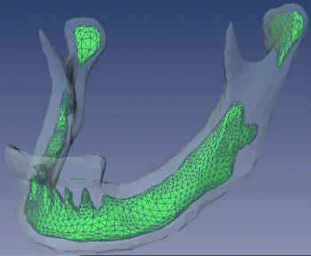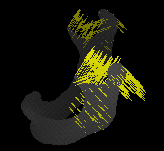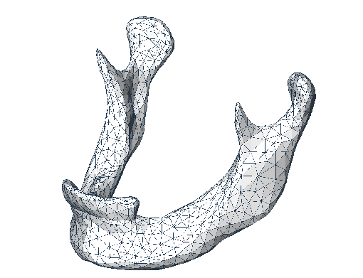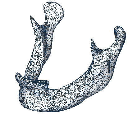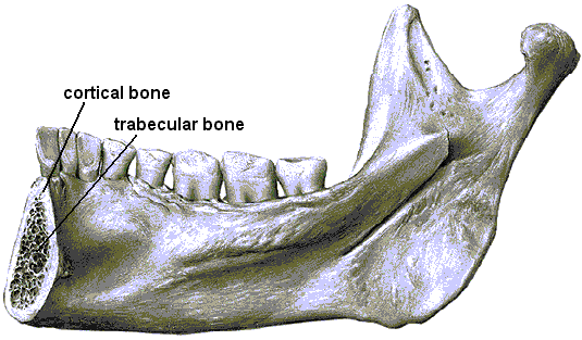
Figure 1: The bone structure of the human mandible.
| Responsible: | Cornelia Kober, Bodo Erdmann, Jens Lang, Peter Deuflhard |
| Cooperation: | R. Sader, H.-F. Zeilhofer,
Technische Universit&t, Klinikum Rechts der Isar, München |
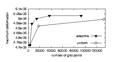
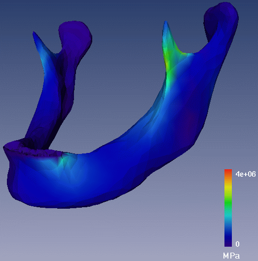
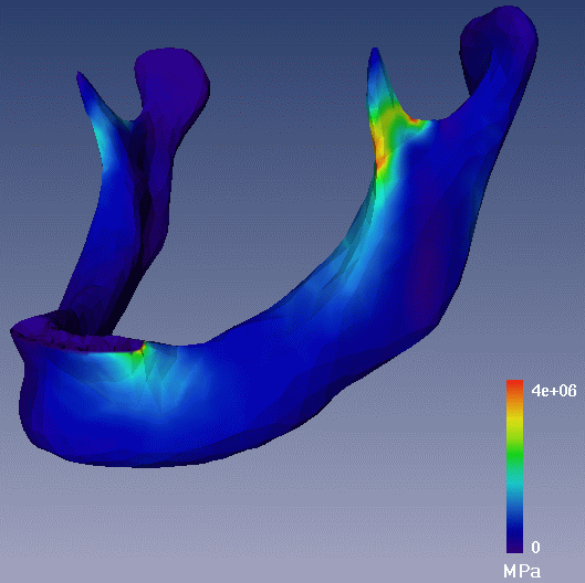
| Literature: | B. Erdmann, C. Kober, J. Lang, P. Deuflhard, H.-F. Zeilhofer, R. Sader, Efficient and Reliable Finite Element Methods for Simulation of the Human Mandible, Report ZIB 01-14, Konrad-Zuse-Zentrum für Informationstechnik Berlin, 2001. |