In biological research, microscopy is a powerful tool to investigate morphological changes in tissues and cell cultures. Modern setups are able to produce 100000 and more images per day with increasing image quality. The images need to be stored, annotated and analyzed. This massive amount of microscopy data is too large to be analyzed manually. There is a clear need for automatic software-based analysis. This project aims to provide a collaborative platform for scientists to work with large collections of microscopy images and assist them with state-of-the-art analysis algorithms. While research questions are evolving, the analyses need to evolve as well. Whereas early applications of microscopy image analysis were simple cell counts, today phenotypes are expected to be detected automatically with machine learning approaches. We hope to achieve best results by implementing a collaborative platform on which biologists and computer scientists work together. It will provide a full pipeline from intuitive data management to the analysis and computation of large data sets. Managing many large data sets together will also enable us to gather meta data and perform more powerful analysis in a bigger context. Collaboration will not only happen between biologists and computer scientists. Data can also be shared between biologists or even published, for example, to provide supplemental data to a journal publication. The developers of the analyses can furthermore collaborate on code and manage the code for the analysis modules.
Publications

2019
Serial synapse formation through filopodial competition for synaptic seeding factors Developmental Cell, 50(4), pp. 447-461, 2019 (preprint available as ZIB-Report 19-45) Mehmet Neset Ozel, Abhishek Kulkarni, Amr Hasan, Josephine Brummer, Marian Moldenhauer, Ilsa-Maria Daumann, Heike Wolfenberg, Vincent J. Dercksen, Ferdi Ridvan Kiral, Martin Weiser, Steffen Prohaska, Max von Kleist, Peter Robin Hiesinger PDF (ZIB-Report)
BibTeX
DOI
Collaborative Data Analysis for Microscopy
2018
Live Observation of Two Parallel Membrane Degradation Pathways at Axon Terminals Current Biology, 28(7), pp. 1027-1038.e4, 2018 Eugene Jennifer Jin, Ferdi Ridvan Kiral, Mehmet Neset Ozel, Lara Sophie Burchardt, Marc Osterland, Daniel Epstein, Heike Wolfenberg, Steffen Prohaska, Peter Robin Hiesinger BibTeX
DOI
Collaborative Data Analysis for Microscopy
2017
Role of bone morphogenetic proteins in sprouting angiogenesis: differential BMP receptor-dependent signaling pathways balance stalk vs. tip cell competence FASEB Journal, 31(11), pp. 4720-4733, 2017 Andreas Benn, Christian Hiepen, Marc Osterland, Christof Schütte, An Zwijsen, Petra Knaus BibTeX
DOI
Collaborative Data Analysis for Microscopy
The role of titanium surface nanotopography on preosteoblast morphology, adhesion and migration Advanced Healthcare Materials, 2017 (preprint available as ZIB-Report 17-06) Yulia Zhukova, Christian Hiepen, Petra Knaus, Marc Osterland, Steffen Prohaska, John W. C. Dunlop, Peter Fratzl, Ekaterina V. Skorb PDF (ZIB-Report)
BibTeX
DOI
Collaborative Data Analysis for Microscopy
2015
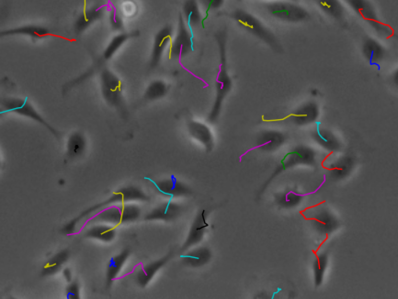
Single Cell Tracking in Phase-Contrast Microscopy EMBL Symposium 2015 - Seeing is Believing - Imaging the Processes of Life, 2015 Marc Osterland, Andreas Benn, Steffen Prohaska, Christof Schütte BibTeX
Collaborative Data Analysis for Microscopy