The analysis of complex molecular structures plays an important role in biochemistry, biophysics, molecular biology and pharmaceutics. Therefore, many visualization techniques have been developed for depicting and interactively analyzing molecules. In this project we address two related problems in molecular analysis:

(i) characterization and visualization of steric hindrance or spatial accessibility of molecules and
(ii) computation and depiction of long molecular paths.

Steric effects in molecules arise from the fact that each atom occupies a certain amount of space. Bringing atoms too close together, requires a high amount of energy due to the Pauli repulsion between overlapping electron clouds. The spatial accessibility of molecules (approximated by sets of spherical atoms) is usually visualized by molecular surfaces. We develop improved and accelerated algorithms for computing such molecular surfaces. Furthermore, we propose a new type of molecular surface that better characterizes the mutual spatial accessibility of molecules.

In molecular dynamics (MD) computations it is difficult to bridge the gap between the fs time scale of the oscillations of covalent bonds and the µs time-scale of the interesting biological processes. For instance, often it is computationally too expensive to compute tracks of substrates to binding sites of proteins. We address this problem by computing sets of geometrically correct molecular paths in the presence of steric hindrances resulting from the van-der-Waals radii of the atoms. These sets of geometrically possible paths constrain the subsets of dynamically possible physical paths. With specialized path visualization techniques, we achieve fast and perceptually optimized interactive representations.

Furthermore, we develop visual analytics tools for uncovering possible molecular paths in complex, dynamic protein configurations. This allows us to analyze the paths of small molecules through transport proteins in cell membranes that become possible due to conformational changes.

Introduction

The analysis and visualization of molecular structures is a large research area. In our project, we focus on methods based on the van-der-Waals spheres of the atoms. We developed fast computation and rendering methods for molecular surfaces. These surfaces are most often used to study molecular behaviour and interactions. They show regions accessible for other molecules like substrates. Furthermore, we work on the extraction of geometric molecular paths. We developed a fast approach to compute all of these paths inside a molecule. Our novel visualization techniques support the analysis and offer easy views into cavities and tunnels.

Surface Definitions

Two very important molecular surfaces are the solvent excluded surface (SES) and the molecular skin surface (MSS). Both surfaces are based on the van-der-Waals spheres of the atoms. The SES is defined as the surface enclosing all points, that are not accessible to a given probe sphere. This probe typically approximates solvents, most often water is used. The SES can be seen as the track of the probe which rolls over the atom spheres. In contrast, the MSS has no chemical background and is defined by a shrink factor which decomposes the surface into quadric patches. If the shrink factor is close to 0, the surface becomes nearly the convex hull of the atom spheres. For a shrink factor equal to 1, the surface is identical to the surface described by the atom spheres, called van-der-Waals surface. The MSS is complete tangent continuous for a shrink factor smaller than 1. In contrast, the SES can have singularities represented by sharp edges and consists of spherical patches as well as toroidal patches of degree four.

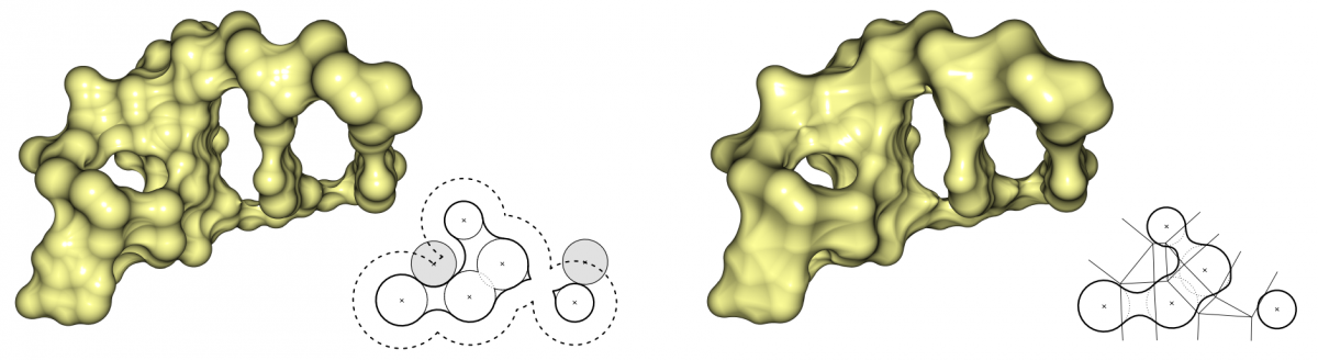
Comparison of SES (left) and MSS (right). The SES is defined by the probe sphere, which approximates a water molecule. For the MSS, we used a shrink factor of 0.3, which creates a topologically similar surface to the SES.

Surface Computation

The analytical description of the SES as well as the MSS can be computed very fast by weighted Voronoi diagrams given by the position of the atoms and their van-der-Waals radii. It is not necessary to compute the correct Voronoi diagram, but an approximation of the Voronoi cells is sufficient, where each cell depends only on the neighboring atoms. Varshney et al. (1994) presented a definition for the neighborhood of atoms for the SES and a fast parallel algorithm. We developed the neighborhood definition for the MSS to use the same concept.

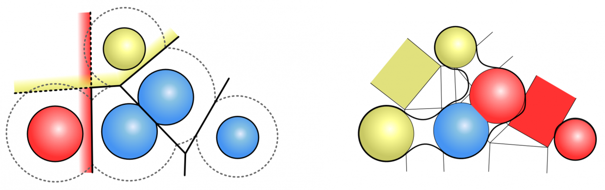
 Approximation of the Voronoi cells of the SES for the red and yellow atom (left). Neighbourhood definition for the MSS (right). The red atoms are neighbours, while the yellow atoms are not.

Surface Rendering

Most visualization tools triangulate the molecular surfaces. This takes several seconds for molecules with a few thousand atoms. For large molecules, the surfaces can consist of several million triangles, so it is not possible to visualize them interactively. We developed a GPU-based raycasting technique for both surfaces, similar to Chavent et al. (2009) and Krone et al (2010). This technique allows the user to visualize surfaces with about 100.000 atoms at interactive frame rates. Combined with our fast computation we are able to render dynamic surfaces for molecules with several thousand atoms. We implemented also classical visualization techniques like coloring, silhouettes, depth darkening and surface blending.

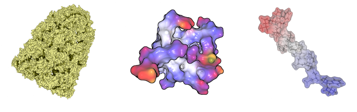
Visualization techniques for molecular surfaces. Depth darkening (left), coloring and silhouettes (middle) and surface blending (right).

Path Computation

Currently, we compute molecular paths based on the geometry of the van-der-Waals spheres of the atoms. The edges of the Voronoi diagram of the atom spheres consists of all possible paths with maximal circular cross section. We developed data structures for a very fast computation of the Voronoi diagram of spheres with the edge tracing algorithm by Kim et al. (2005). A lot of paths of the Voronoi diagram are redundant, because there are more significant paths. Furthermore, there are many paths outside of the domain of the molecule and paths with too small or negative cross sections. We developed a novel filtering technique to keep only the most significant paths of the molecule. The overall computation takes less than one minute, even for molecules with approximately 100.000 atoms.


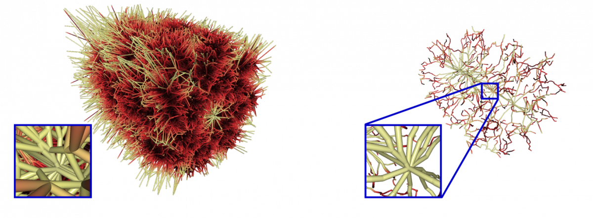
Voronoi diagram of the chaperonin protein 1AON clipped with the bounding box and after filtering.

Path Visualization

Path visualization in dense geometry is a challenging problem in computer graphics. We developed several techniques to enable a fast analysis of the molecular structure. In a first step, we place a lot of small point lights along the extracted paths, which highlight the molecular surface in cavities and tunnels. To keep interactive frame rates, we use deferred shading with high dynamic range rendering to avoid artifacts. We also implemented screen space ambient occlusion for a better depth impression. Furthermore, we provide surface clipping for selected paths, which allows one to analyze the dimension and the cavities of the selected paths.

Our path lighting with surface clipping for the selected paths (red)

Dynamic Paths

In order to analyze cavities and channels in dynamic molecular data from simulations, we compute all molecular paths for all time steps of the trajectory. The user is allowed to select one or more path components of the overall path graph for an arbitrary time step. Note that each path component corresponds to exactly one cavity or channel. The selected path components will be traced over time by analyzing the intersections of the corresponding cavities with all cavities in the consecutive time step. A valid tracing is found if an intersection is large enough so that a given probe sphere is able to move from one cavity into a cavity of the consecutive time step. During this analysis, cavity splits and merges are detected and visualized in a 2-dimensional graph. Each path in this graph describes a possible geometrical dynamic behavior of the cavity. Thus, dynamic molecular channels can be detected. The dynamic cavities and channels can be visualized either by the skin surface or volume rendering. We tested our tool for the membrane protein bacteriorhodopsin, which works as a light driven proton pump.

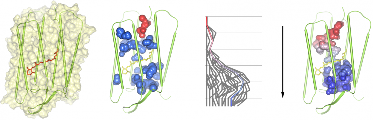
(1) A bacteriorhodopsin monomer displayed as surface and cartoons, with the retinal, shown as bonds, covalently bound to Lys216. (2) All cavities of the monomer as found at time step 651; the cavity shown in red is traced over time. (3) Split and merge graph of path tracing. (4) A dynamic molecular path, connecting the cytoplasmic (top) and the extracellular side (bottom), based on the selection in the split and merge graph.

Publications

2019
Interactive Visualization of RNA and DNA Structures IEEE Transactions on Visualization and Computer Graphics, 25(1), pp. 967-976, 2019 (preprint available as ZIB-Report 18-33) Norbert Lindow, Daniel Baum, Morgan Leborgne, Hans-Christian Hege PDF (ZIB-Report)
BibTeX
DOI
Analysis and Visualization of Molecular Structures
2018
A multiscale perturbation expansion approach for Markov state modeling of non-stationary molecular dynamics SIAM J. Multiscale Model. Simul., 16(4), pp. 1455-1485, 2018 (preprint available as ) Péter Koltai, Christof Schütte BibTeX
DOI
Analysis and Visualization of Molecular Structures
Atomic Accessibility Radii for Molecular Dynamics Analysis Workshop on Molecular Graphics and Visual Analysis of Molecular Data, 2018, ISBN: 978-3-03868-061-1 (preprint available as ZIB-Report 18-18) Norbert Lindow, Daniel Baum, Hans-Christian Hege PDF (ZIB-Report)
BibTeX
DOI
Analysis and Visualization of Molecular Structures
MSM/RD: Coupling Markov state models of molecular kinetics with reaction-diffusion simulations Journal of Chemical Physics, 148(214107), 2018 Manuel Dibak, Mauricio J. del Razo, David De Sancho, Christof Schütte, Frank Noé BibTeX
DOI
Analysis and Visualization of Molecular Structures
2016
Visual Analysis of Atomic Structures Based on the Hard-Sphere Model Doctoral thesis, Freie Universität Berlin, Christof Schütte (Advisor), 2016 Norbert Lindow PDF
BibTeX
URN
Analysis and Visualization of Molecular Structures
Visual Analysis of Biomolecular Cavities: State of the Art Computer Graphics Forum, 35(3), pp. 527-551, 2016 (preprint available as ZIB-Report 16-42) Michael Krone, Barbora Kozlíková, Norbert Lindow, Marc Baaden, Daniel Baum, Julius Parulek, Hans-Christian Hege, Ivan Viola PDF (ZIB-Report)
BibTeX
DOI
Analysis and Visualization of Molecular Structures
Visualization of Biomolecular Structures: State of the Art Revisited Computer Graphics Forum, 36(8), pp. 178-204, 2016 Barbora Kozlíková, Michael Krone, Martin Falk, Norbert Lindow, Marc Baaden, Daniel Baum, Ivan Viola, Julius Parulek, Hans-Christian Hege BibTeX
DOI
Analysis and Visualization of Molecular Structures
2015
Membrane Protein Structure, Function and Dynamics: A Perspective from Experiments and Theory Journal of Membrane Biology, 248(4), pp. 611-640, 2015 Zoe Cournia, Toby W. Allen, Ioan Andricioaei, Bruno Antonny, Daniel Baum, Grace Brannigan, Nicolae-Viorel Buchete, Jason T. Deckman, Lucie Delemotte, Coral del Val, Ran Friedman, Paraskevi Gkeka, Hans-Christian Hege, Jérôme Hénin, Marina A. Kasimova, Antonios Kolocouris, Michael L. Klein, Syma Khalid, Joanne Lemieux, Norbert Lindow, Mahua Roy, Jana Selent, Mounir Tarek, Florentina Tofoleanu, Stefano Vanni, Sinisa Urban, David J. Wales, Jeremy C. Smith, Ana-Nicoleta Bondar BibTeX
DOI
Analysis and Visualization of Molecular Structures
Visualization of Biomolecular Structures: State of the Art EuroVis 2015 STARS Proceedings, pp. 61-81, 2015 (preprint available as ZIB-Report 15-63) Barbora Kozlikova, Michael Krone, Norbert Lindow, Martin Falk, Marc Baaden, Daniel Baum, Ivan Viola, Julius Parulek, Hans-Christian Hege PDF (ZIB-Report)
BibTeX
DOI
Analysis and Visualization of Molecular Structures
2014
Ligand Excluded Surface: A New Type of Molecular Surface IEEE Transactions on Visualization and Computer Graphics, 20(12), pp. 2486-2495, 2014 (preprint available as ZIB-Report 14-27) Norbert Lindow, Daniel Baum, Hans-Christian Hege PDF (ZIB-Report)
BibTeX
DOI
Analysis and Visualization of Molecular Structures
Visual analysis of molecular dynamics data using geometric and topological methods Forum "Math for Industry" 2014, pp. 45-46, Vol.57, MI Lecture Notes, Kyushu University, 2014 Hans-Christian Hege BibTeX
Analysis and Visualization of Molecular Structures
2013
Exploring cavity dynamics in biomolecular systems BMC Bioinformatics, Vol.14, 2013 Norbert Lindow, Daniel Baum, Ana-Nicoleta Bondar, Hans-Christian Hege BibTeX
DOI
Analysis and Visualization of Molecular Structures
2012
Dynamic Channels in Biomolecular Systems: Path Analysis and Visualization Proceedings of IEEE Symposium on Biological Data Visualization (biovis’12), pp. 99-106, 2012 Norbert Lindow, Daniel Baum, Ana-Nicoleta Bondar, Hans-Christian Hege BibTeX
DOI
Analysis and Visualization of Molecular Structures
2011
Voronoi-Based Extraction and Visualization of Molecular Paths IEEE Transactions on Visualization and Computer Graphics, 17(12), pp. 2025-2034, 2011 Norbert Lindow, Daniel Baum, Hans-Christian Hege BibTeX
DOI
Analysis and Visualization of Molecular Structures
2010
Accelerated Visualization of Dynamic Molecular Surfaces Comput. Graph. Forum, Vol.29, pp. 943-952, 2010 Norbert Lindow, Daniel Baum, Steffen Prohaska, Hans-Christian Hege BibTeX
DOI
Analysis and Visualization of Molecular Structures