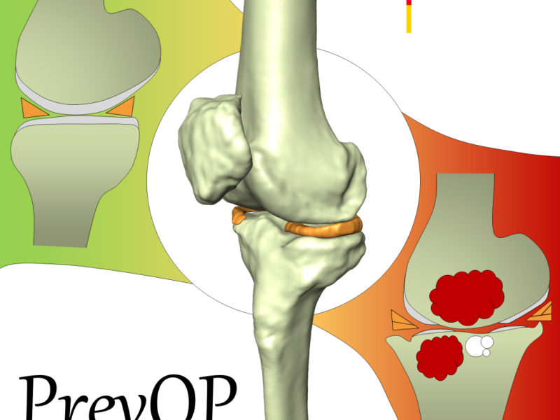
Within the PrevOP research network "Preventing the progression of primary Osteoarthritis by high impact long-term Physical exercise regimen – key mechanisms, efficacy, and long-term results." the aim of sub-project 4 is to assess and to quantitatively analyze morphological and structural changes related to osteoarthritis (OA) with respect to different levels of exercise. In arthritic knees typical OA-related findings are the degeneration of cartilage, menisci, and ligaments. Furthermore, in the majority of cases of severe OA, bone marrow lesions and subarticular cysts occur. It is assumed that these structures can be quantitatively assessed with medical imaging techniques, in particular MRI (Fig. 1). In this project, using experts' reading and machine learning techniques, OA-related quantitative image features will be identified for every relevant structure. Combining all image features might lead to a novel image-based OA-score. This OA-score could be used to support the hypothesis that exercise slows down the progression of OA.

PrevOp OA features

Fig 1: Features of osteoarthritis: Cartilage defects, meniscal tears, bone marrow lesions and subarticular cysts.

Identifying OA-related features with data of the Osteoarthritis Initiative (OAI) database

In the OAI database MRIs and radiographs are publicly available for almost 5.000 patients. In addition to basic scoring data (i.e. WOMAC and Kellgren-Lawrence score) several image assessment studies have been carried out for OAI data:

  • Whole-Organ Magnetic Resonance Imaging Score (WORMS) and Boston Leeds Osteoarthritis Knee Score (BLOKS) scoring results have been published in Mai 2011 for 115 patients
  • MRI Osteoarthritis Knee Score (MOAKS) reading results have been published in April 2015 for 600 patients

In this project, image features will be extracted for each OA-related structure (cartilage, menisci, ligaments, bone marrow lesions and subarticular cysts). Employing methods of machine learning and using the data of the WORMS, BLOKS and MOAKS image assessment studies, the correlation between these image features and the experts' reading will be investigated. Features, which were identified as being essential for OA progression, will be combined to an image-based OA score (Fig. 2).
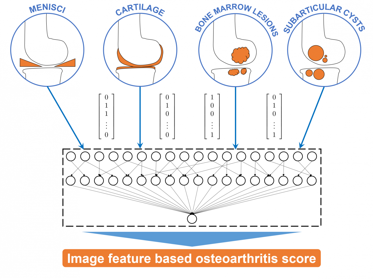
Combining image feature based measurements to an OA score

Fig 2: Image-based quantitative features are extracted for the cartilage, menisci, bone marrow lesions and subarticular cysts. These image features are combined to a novel osteoarthritis score.

Automated segmentation of knee bones, cartilage, and menisci

To generate specific regions of interest (ROI) for the considered anatomical structures in the MRI data, the knee bones (femur and tibia), the femoral and tibial cartilage [1], and the menisci [2] are automatically segmented using methods based on convolutional neural networks and statistical shape models.

Analysis of the knee menisci

Using the 3D segmentations of the menisci and the knee bones quantitative measurements (meniscal volume, meniscal extrusion, and tibial coverage) were calculated [2]. We investigated the correlation of these measurements and the OA grade using data from the OAI database [2].

Also, we pursued the aim to detect meniscal tears in MRI data by using deep learning methods with the automatically determined meniscal ROI and MOAKS semi-quantitative meniscal morphology reading [3].

Analysis of the knee bones and cartilage

An deep learning based method for the automated segmentation of knee bones and cartilage was developed and thoroughly validated [1]. We will investigate the usability of cartilage volume and thickness measures derived from these segmentations as suitable and reliable OA biomarkers. We employed a multi-loss function to further increase the accuracy of cartilage volume measurements [4].

Analysis of the knee alignment

A deep learning based method for the automated assessment of knee alignment from full-leg X-Ray images was developed [5]. The accuracy was evaluated for over 3,000 datasets from the OAI database.

Data availability

All generated data are available at pubdata.zib.de.

References

[1] F Ambellan, A Tack, M Ehlke, S Zachow (2019). Automated Segmentation of Knee Bone and Cartilage combining Statistical Shape Knowledge and Convolutional Neural Networks: Data from the Osteoarthritis Initiative. Medical Image Analysis; 52(2), p. 109--118

[2] A Tack, A Mukhopadhyay, S Zachow (2018). Knee Menisci Segmentation using Convolutional Neural Networks: Data from the Osteoarthritis Initiative. Osteoarthritis and Cartilage; 26(5), p. 680--688.

[3] V Reddy, A Tack, B Preim, S Zachow (2017). Automatic Classification of 3D MRI data using Deep Convolutional Neural Networks. Masters thesis, Otto-von-Guericke-Universität Magdeburg

[4] A Tack, S Zachow (2019). Accurate Automated Volumetry of Cartilage of the Knee using Convolutional Neural Networks: Data from the Osteoarthritis Initiative. ISBI 2019

[5] A Tack, B Preim, S Zachow (2021). Fully automated Assessment of Knee Alignment from Full-Leg X-Rays employing a "YOLOv4 And Resnet Landmark regression Algorithm" (YARLA): Data from the Osteoarthritis Initiative. Computer Methods and Programs in Biomedicine; epub ahead of print


Publications

2024
Machine Learning-based Assessment of Multiple Anatomical Structures in Medical Image Data for Diagnosis and Prediction of Knee Osteoarthritis Doctoral thesis, Technische Universität Berlin, Stefan Zachow (Advisor), 2024 Alexander Tack BibTeX
DOI
Analysis and quantification of morphological and structural changes in cartilage
2021
A Deep Learning Method for Automated Detection of Meniscal Tears in Meniscal Sub-Regions in 3D MRI Data Master's thesis, Freie Universität Berlin, Tack Alexander, Zachow Stefan (Advisors), 2021 Alexey Shestakov BibTeX
Analysis and quantification of morphological and structural changes in cartilage
A deep multi-task learning method for detection of meniscal tears in MRI data from the Osteoarthritis Initiative database Frontiers in Bioengineering and Biotechnology, section Biomechanics, pp. 28-41, 2021 (preprint available as ZIB-Report 21-33) Alexander Tack, Alexey Shestakov, David Lüdke, Stefan Zachow PDF (ZIB-Report)
BibTeX
DOI
Analysis and quantification of morphological and structural changes in cartilage
Fully automated Assessment of Knee Alignment from Full-Leg X-Rays employing a "YOLOv4 And Resnet Landmark regression Algorithm" (YARLA): Data from the Osteoarthritis Initiative Computer Methods and Programs in Biomedicine, 205(106080), 2021 Alexander Tack, Bernhard Preim, Stefan Zachow PDF
BibTeX
DOI
Analysis and quantification of morphological and structural changes in cartilage
Investigation of Options to Handle 3D MRI Data via Convolutional Neural Networks Application in Knee Osteoarthritits Classification Master's thesis, Otto-von-Guericke-Universität Magdeburg, Alexander Tack, Stefan Zachow (Advisors), 2021 Jan Krause BibTeX
Analysis and quantification of morphological and structural changes in cartilage
Towards novel osteoarthritis biomarkers: Multi-criteria evaluation of 46,996 segmented knee MRI data from the Osteoarthritis Initiative PLOS One, 16(10), 2021 Alexander Tack, Felix Ambellan, Stefan Zachow BibTeX
DOI
Analysis and quantification of morphological and structural changes in cartilage
Towards novel osteoarthritis biomarkers: Multi-criteria evaluation of 46,996 segmented knee MRI data from the Osteoarthritis Initiative (Supplementary Material) PLOS One, 16(10), 2021 Alexander Tack, Felix Ambellan, Stefan Zachow BibTeX
DOI
Analysis and quantification of morphological and structural changes in cartilage
Unsupervised Detection of Disturbances in 2D Radiographs 2021 IEEE 18th International Symposium on Biomedical Imaging (ISBI), pp. 367-370, 2021 Laura Estacio, Moritz Ehlke, Alexander Tack, Eveling Castro-Gutierrez, Hans Lamecker, Rensso Mora, Stefan Zachow BibTeX
DOI
Analysis and quantification of morphological and structural changes in cartilage
2019
Accurate Automated Volumetry of Cartilage of the Knee using Convolutional Neural Networks: Data from the Osteoarthritis Initiative IEEE 16th International Symposium on Biomedical Imaging (ISBI 2019), pp. 40-43, 2019 (preprint available as ZIB-Report 19-05) Alexander Tack, Stefan Zachow PDF (ZIB-Report)
BibTeX
DOI
Analysis and quantification of morphological and structural changes in cartilage
Automated Hip Knee Ankle Angle Determination using Convolutional Neural Networks Master's thesis, Otto-von-Guericke-Universität Magdeburg, Felix Ambellan, Alexander Tack, Stefan Zachow (Advisors), 2019 Henok Hagos Gidey PDF
BibTeX
URN
Analysis and quantification of morphological and structural changes in cartilage
Automated Segmentation of Knee Bone and Cartilage combining Statistical Shape Knowledge and Convolutional Neural Networks: Data from the Osteoarthritis Initiative Medical Image Analysis, 52(2), pp. 109-118, 2019 (preprint available as ZIB-Report 19-06) Felix Ambellan, Alexander Tack, Moritz Ehlke, Stefan Zachow PDF (ZIB-Report)
BibTeX
DOI
Analysis and quantification of morphological and structural changes in cartilage
Automated Segmentation of Knee Bone and Cartilage combining Statistical Shape Knowledge and Convolutional Neural Networks: Data from the Osteoarthritis Initiative (Supplementary Material) Medical Image Analysis, 52(2), pp. 109-118, 2019 (OAI-ZIB dataset) Felix Ambellan, Alexander Tack, Moritz Ehlke, Stefan Zachow BibTeX
DOI
Analysis and quantification of morphological and structural changes in cartilage
Comparison of 2D and 3D CNNs for Classification of Knee MRI Bachelor's thesis, Freie Universität Berlin, Alexander Tack, Felix Ambellan (Advisors), 2019 Mona Prendke PDF
BibTeX
URN
Analysis and quantification of morphological and structural changes in cartilage
Statistical Shape Models - Understanding and Mastering Variation in Anatomy Biomedical Visualisation, Paul M. Rea (Ed.), Springer Nature Switzerland AG, 1, pp. 67-84, 2019, ISBN: 978-3-030-19384-3, ISBN: 978-3-030-19385-0 (preprint available as ZIB-Report 19-13) Felix Ambellan, Hans Lamecker, Christoph von Tycowicz, Stefan Zachow PDF (ZIB-Report)
BibTeX
DOI
Analysis and quantification of morphological and structural changes in cartilage
2018
Automated Segmentation of Knee Bone and Cartilage combining Statistical Shape Knowledge and Convolutional Neural Networks: Data from the Osteoarthritis Initiative Medical Imaging with Deep Learning, 2018 Felix Ambellan, Alexander Tack, Moritz Ehlke, Stefan Zachow PDF
BibTeX
Analysis and quantification of morphological and structural changes in cartilage
Knee Menisci Segmentation using Convolutional Neural Networks: Data from the Osteoarthritis Initiative Osteoarthritis and Cartilage, 26(5), pp. 680-688, 2018 (preprint available as ZIB-Report 18-15) Alexander Tack, Anirban Mukhopadhyay, Stefan Zachow PDF (ZIB-Report)
BibTeX
DOI
Analysis and quantification of morphological and structural changes in cartilage
Knee Menisci Segmentation using Convolutional Neural Networks: Data from the Osteoarthritis Initiative (Supplementary Material) 2018 Alexander Tack, Anirban Mukhopadhyay, Stefan Zachow BibTeX
DOI
Analysis and quantification of morphological and structural changes in cartilage
2017
Automatic Classification of 3D MRI data using Deep Convolutional Neural Networks Master's thesis, Otto-von-Guericke-Universität Magdeburg, Stefan Zachow, Alexander Tack (Advisors), 2017 Gutha Vaishnavi Reddy BibTeX
Analysis and quantification of morphological and structural changes in cartilage
Evaluating two methods for Geometry Reconstruction from Sparse Surgical Navigation Data Proceedings of the Jahrestagung der Deutschen Gesellschaft für Computer- und Roboterassistierte Chirurgie (CURAC), pp. 24-30, Vol.16, 2017 (preprint available as ZIB-Report 17-71) Felix Ambellan, Alexander Tack, Dave Wilson, Carolyn Anglin, Hans Lamecker, Stefan Zachow PDF (ZIB-Report)
BibTeX
URN
Analysis and quantification of morphological and structural changes in cartilage
Shape-aware Surface Reconstruction from Sparse 3D Point-Clouds Medical Image Analysis, Vol.38, pp. 77-89, 2017 Florian Bernard, Luis Salamanca, Johan Thunberg, Alexander Tack, Dennis Jentsch, Hans Lamecker, Stefan Zachow, Frank Hertel, Jorge Goncalves, Peter Gemmar PDF
BibTeX
DOI
Analysis and quantification of morphological and structural changes in cartilage
Validation of three-dimensional models of the distal femur created from surgical navigation point cloud data for intraoperative and postoperative analysis of total knee arthroplasty International Journal of Computer Assisted Radiology and Surgery, 12(12), pp. 2097-2105, 2017 David Wilson, Carolyn Anglin, Felix Ambellan, Carl Martin Grewe, Alexander Tack, Hans Lamecker, Michael Dunbar, Stefan Zachow PDF
BibTeX
DOI
Analysis and quantification of morphological and structural changes in cartilage
2016
Shape-aware Surface Reconstruction from Sparse Data arXiv, p. 1602.08425v1, 2016 Florian Bernard, Luis Salamanca, Johan Thunberg, Alexander Tack, Dennis Jentsch, Hans Lamecker, Stefan Zachow, Frank Hertel, Jorge Goncalves, Peter Gemmar PDF
BibTeX
arXiv
Analysis and quantification of morphological and structural changes in cartilage
2015
Bewegungsfeldschätzung in artefaktbehafteten 4D-CT-Bilddaten: Vergleich von paar- und gruppenweiser Registrierung 21st Annual Meeting of the German-Society-for-Radiation-Oncology, Vol.Supplement 1, pp. 65-65, Springer: Strahlentherapie und Onkologie, 191, 2015 Alexander Tack, Yuske Kobayashi, Tobias Gauer, Alexander Schlaefer, René Werner PDF
BibTeX
DOI
Analysis and quantification of morphological and structural changes in cartilage
Groupwise Registration for Robust Motion Field Estimation in Artifact-Affected 4D CT Images ICART: Imaging and Computer Assistance in Radiation Therapy: A workshop held on Friday 9th October as part of MICCAI 2015 in Munich, Germany. MICCAI workshop. 2015., pp. 18-25, 2015 Alexander Tack, Yuske Kobayashi, Tobias Gauer, Alexander Schlaefer, René Werner PDF
BibTeX
Analysis and quantification of morphological and structural changes in cartilage
Gruppenweise Registrierung zur robusten Bewegungsfeldschätzung in artefaktbehafteten 4D-CT-Bilddaten Master's thesis, Technische Universität Hamburg-Harburg, Alexander Schlaefer, Michael Morlock, René Werner (Advisors), 2015 Alexander Tack BibTeX
Analysis and quantification of morphological and structural changes in cartilage