The aim of this project is the development of MRI-based methods and technologies for non-invasive in vivo assessment of mechanical loads in soft tissue structures in knee joints. Such an in vivo assessment of 3D soft tissue kinematics, together with their incident loads, would pave the way for a better understanding of deformations of tendons, ligaments and menisci in healthy subjects as well as their changes due to pathologies (e.g., after ruptures of the anterior cruciate ligament in young vs. elderly patients).
PROJECT GOALS
Given the short acquisition times available in the dynamic imaging setup only partial information (low-resolution images) can be acquired. Therefore, specialized reconstruction methods have to be developed in order to extract the motion of soft tissues from the incomplete data by correlating the information from high-resolution static images with low-resolution dynamic sequences.
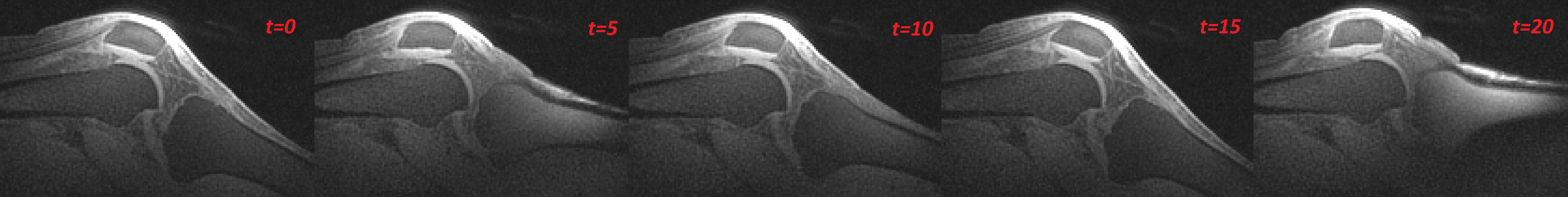
Selected time frames of the dynamic MRI sequence show large changes of image intensity.
DYNAMIC AND STATIC MRI DATA
Ultra-short echo-time (UTE) MRI sequences have been developed at Jena University Hospital, made specifically for dynamic imaging of human knee joints. Based on these sequences and a custom-made device for guided knee motion, developed at Charitè, high-resolution static and low-resolution dynamic MRI data was acquired.
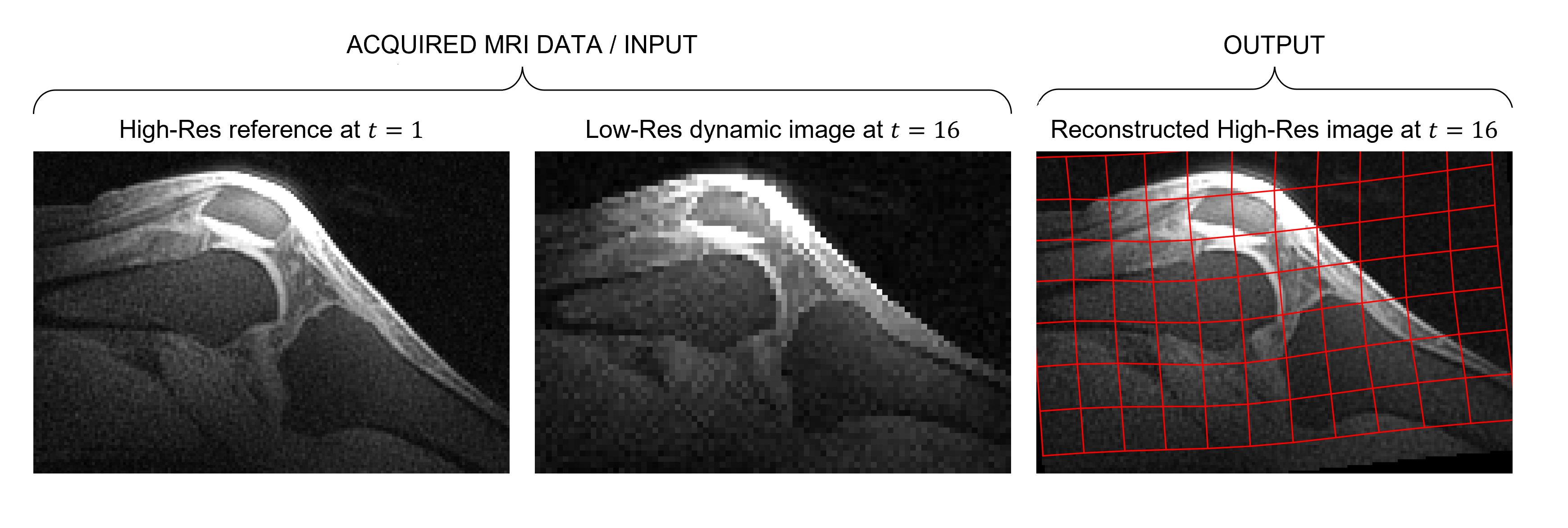
Acquired MRI knee data and its reconstruction. The reconstructed image is overlaid with a grid which represents how the HR reference image was deformed to align with corresponding LR dynamic time frame.
LOG-EUCLIDEAN REGISTRATION OF DYNAMIC MR IMAGES
As an alternative to non-linear B-Spline-based registration, a log-euclidean polyaffine registration framework is investigated. In this framework, each bone is individually registered between adjacent time frames in a rigid manner. In the second step, the resulting transformations are combined using the exponential weighting function and the log-euclidean mapping to obtain the final reconstructed image and the deformation field.
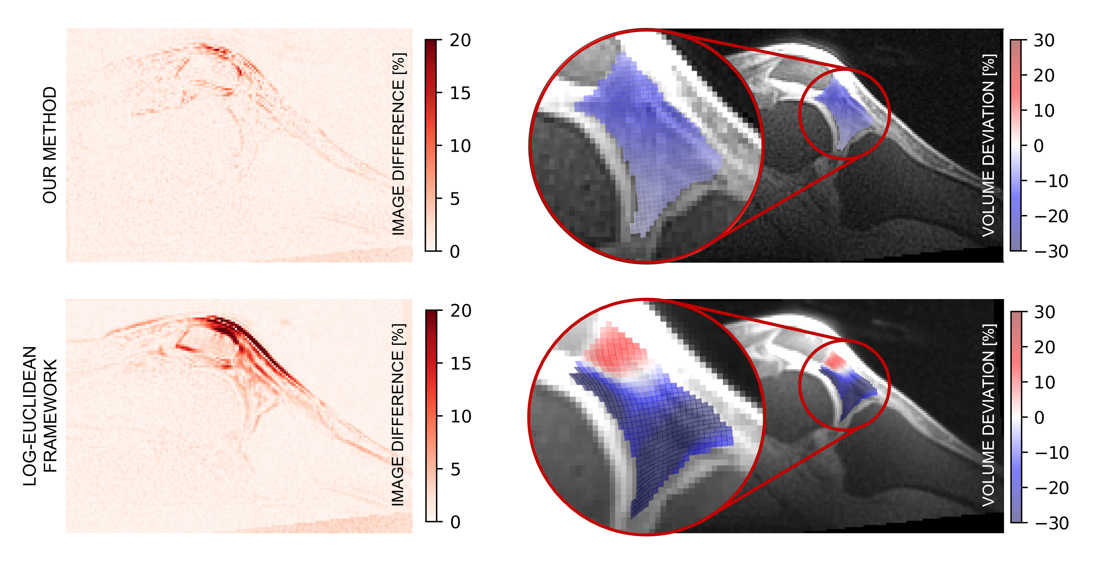
Comparison between our B-spline-based registration approach and the log-euclidean polyaffine framework for a single time frame. Slices on the left show the normalized image difference between the HR reconstruction and the HR ground truth, while images on the right show the relative volume change for a 2D slice of Hoffa’s fat pad.
High spatial resolution dynamic MRI
After applying the registration approach, a high spatial resolution dynamic image sequence is obtained. Further tissues deformations during movement can be assessed.
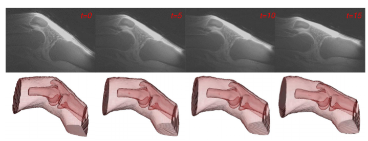
Reconstructed dynamic HR spatial resolution image (top) and dynamic displacement of tissues (bottom)