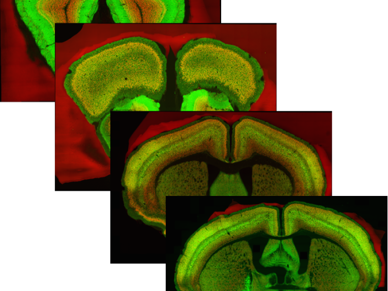
Imaging and analyzing the entire brain of small vertebrates at maximal confocal microscopy resolution seems possible today. One could, for example, derive a complete distribution of neuron cell bodies; screen for unlocalized changes, such as amyloid plaques; or build detailed anatomical reference systems. The goal of this project is to develop an image acquisition and processing pipeline for entire brains and apply it to a mouse brain.
Publications

2011
Large-scale automated histology in the pursuit of connectomes Journal of Neuroscience, 31(45), pp. 16125-16138, 2011 David Kleinfeld, Arjun Bharioke, Pablo Blinder, David Bock, Kevin Briggman, Dmitri Chklovskii, Winfried Denk, Moritz Helmstaedter, John Kaufhold, Wei-Chung Lee, Hanno Meyer, Kristina Micheva, Marcel Oberlaender, Steffen Prohaska, R. Reid, Stephen Smith, Shinya Takemura, Philbert Tsai, Bert Sakmann BibTeX
DOI
Confocal Microscopy for an Entire Mouse Brain at Cellular Resolution