Unlike most other vertebrate animals, sharks and rays have skeletons made entirely of cartilage and not bone. In contrast to the cartilage in our knees, however, a shark’s skeleton is “tessellated”: skeletal elements made of uncalcified cartilage are wrapped in an outer layer of thousands of mineralized tiles called tesserae. Although this character has defined the shark and ray lineage for hundreds of millions of years, it is still unclear what role skeletal tiling plays in the mechanics and growth of this ancient, composite skeletal material. The HFSP-funded project aims to describe the tiling morphologies of shark and ray cartilaginous skeletons, to understand their importance to the mechanical behavior of the skeleton. Through several collaborations, we use an integrative and iterative approach of morphological analyses, materials and mechanical testing, and modeling (physical, digital and theoretical). The subproject at ZIB focuses on the analysis of µCT data of shark and ray skeletons. In particular, the aim is to develop tools for flexible segmentations of the tessellated network, allowing the first 3d quantifications and visualizations of these tiling patterns and allowing comparison among species and developmental stages, but also with idealized tiled surfaces.

Segmentation Workflow

We have developed a workflow for the segmentation of µCT data sets of ray tesserae. Since the scalar value distribution of the tesserae does not enable a separation of the individual tesserae, we are using the following multi-step approach:

  1. Pre-processing with anisotropic diffusion
  2. Binary segmentation of the tesseral layer using a local thresholding approach
  3. Computation of a 2D distance transform of the tesseral layer
  4. Segmentation of individual tesserae based on hierarchical watershed on the 2D distance map
  5. Computation of a graph representation for comfortable interactive correction of segmentation errors

Segmentation Workflow

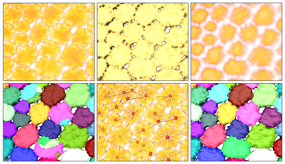
Segmentation workflow (from left to right and top to bottom): Volume rendering of original data; surface of binary segmentation; volume rendering of distance field; initial segmentation using distance field; graph representation; final segmentation.

For evaluation, the segmentation pipeline was applied to the right hyomandibulae of three ages of stingray, each consisting of thousands of tesserae. Quantitative evaluation was done by comparing manually-placed landmarks with the segmented tesserae (via precision/recall values), and by computing RAND and VI values for manually-segmented areas created by four persons.

Results

The segmentation pipeline was applied successfully to the left and right hyomandibulae of six ages of stingray in approximately 5 hours of work per dataset (compared to days or weeks of work for a manual segmentation). This will allow the first quantifications ever done for this kind of biological, tesselated data.

Publications

2023
High-throughput segmentation, data visualization, and analysis of sea star skeletal networks Journal of Structural Biology, 215(2), p. 107955, 2023 Lara Tomholt, Daniel Baum, Robert J. Wood, James C. Weaver BibTeX
DOI
Segmentation and Quantitative Analysis of Cartilaginous Fish Skeletons
2022
Estimating 3D Shape of the Head Skeleton of Basking Sharks Using Annotated Landmarks on a 2D Image Master's thesis, Freie Universität Berlin, Daniel Baum, Christoph von Tycowicz, Mason N. Dean (Advisors), 2022 Martha Paskin PDF
BibTeX
Segmentation and Quantitative Analysis of Cartilaginous Fish Skeletons
Ontogeny of a tessellated surface: carapace growth of the longhorn cowfish Lactoria cornuta Journal of Anatomy, 241(3), pp. 565-580, 2022 Lennart Eigen, Daniel Baum, Mason N. Dean, Daniel Werner, Jan Wölfer, John A. Nyakatura BibTeX
DOI
Segmentation and Quantitative Analysis of Cartilaginous Fish Skeletons
2020
Co-aligned chondrocytes: Zonal morphological variation and structured arrangement of cell lacunae in tessellated cartilage Bone, Vol.134, p. 115264, 2020 (preprint available as ZIB-Report 20-04) Júlia Chaumel, Merlind Schotte, Joseph J. Bizzarro, Paul Zaslansky, Peter Fratzl, Daniel Baum, Mason N. Dean PDF (ZIB-Report)
BibTeX
DOI
Segmentation and Quantitative Analysis of Cartilaginous Fish Skeletons
Image analysis pipeline for segmentation of a biological porosity network, the lacuno-canalicular system in stingray tesserae MethodsX, Vol.7, p. 100905, 2020 (preprint available as ZIB-Report 20-12) Merlind Schotte, Júlia Chaumel, Mason N. Dean, Daniel Baum PDF (ZIB-Report)
BibTeX
DOI
Segmentation and Quantitative Analysis of Cartilaginous Fish Skeletons
2019
High-Throughput Segmentation of Tiled Biological Structures using Random-Walk Distance Transforms Integrative And Comparative Biology, 2019 (preprint available as ZIB-Report 19-33) Daniel Baum, James C. Weaver, Igor Zlotnikov, David Knötel, Lara Tomholt, Mason N. Dean PDF (ZIB-Report)
BibTeX
DOI
Segmentation and Quantitative Analysis of Cartilaginous Fish Skeletons
2018
Ambient occlusion – a powerful algorithm to segment shell and skeletal intrapores in computed tomography data ZIB-Report 18-14 Jürgen Titschack, Daniel Baum, Kei Matsuyama, Karin Boos, Claudia Färber, Wolf-Achim Kahl, Karsten Ehrig, Dietmar Meinel, Carmen Soriano, Stuart R. Stock PDF
BibTeX
URN
Segmentation and Quantitative Analysis of Cartilaginous Fish Skeletons
2017
Automated Segmentation of Complex Patterns in Biological Tissues: Lessons from Stingray Tessellated Cartilage PLOS ONE, 2017 (preprint available as ZIB-Report 17-62) David Knötel, Ronald Seidel, Steffen Prohaska, Mason N. Dean, Daniel Baum PDF (ZIB-Report)
BibTeX
DOI
Segmentation and Quantitative Analysis of Cartilaginous Fish Skeletons
Automated Segmentation of Complex Patterns in Biological Tissues: Lessons from Stingray Tessellated Cartilage (Supplementary Material) 2017 David Knötel, Ronald Seidel, Paul Zaslansky, Steffen Prohaska, Mason N. Dean, Daniel Baum BibTeX
DOI
Segmentation and Quantitative Analysis of Cartilaginous Fish Skeletons
Ultrastructural, material and crystallographic description of endophytic masses – a possible damage response in shark and ray tessellated calcified cartilage Journal of Structural Biology, 2017 Ronald Seidel, Michael Blumer, Paul Zaslansky, David Knötel, Daniel R. Huber, James C. Weaver, Peter Fratzl, Sidney Omelon, Luca Bertinetti, Mason N. Dean BibTeX
DOI
Segmentation and Quantitative Analysis of Cartilaginous Fish Skeletons
2016
Biological strategies for fatique and wear avoidance: lessons from stingray skeletons and teeth Poster, Tomography for Scientific Advancement symposium (ToScA), 2016 Mason N. Dean, Ahmed Hosny, Ronald Seidel, Daniel Baum BibTeX
Segmentation and Quantitative Analysis of Cartilaginous Fish Skeletons
To build a shark: 3D tiling laws of tessellated cartilage Abstract in Integrative and Comparative Biology; conference Society of Integrative and Comparative Biology annual meeting, January 3-7, 2016, Portland, USA, Vol.56 (suppl 1), 2016 Mason N. Dean, R. Seidel, David Knötel, K. Lyons, Daniel Baum, James C. Weaver, Peter Fratzl BibTeX
Segmentation and Quantitative Analysis of Cartilaginous Fish Skeletons
Understanding the Tiling Rules of the Tessellated Mineralized Endoskeleton of Sharks and Rays Poster, Euro Bio-inspired Materials 2016, Potsdam, Germany, February 22 - 25, 2016, 2016 David Knötel, Ronald Seidel, Ahmed Hosny, Paul Zaslansky, James C. Weaver, Daniel Baum, Mason N. Dean BibTeX
Segmentation and Quantitative Analysis of Cartilaginous Fish Skeletons
2015
Automatische Dickenbestimmung der mineralisierten Schicht in Skelettelementen von Knorpelfischen anhand von CT- Bilddaten Bachelor's thesis, Beuth Hochschule für Technik Berlin, Daniel Baum, Ute Wagner (Advisors), 2015 Merlind Schotte BibTeX
Segmentation and Quantitative Analysis of Cartilaginous Fish Skeletons
Segmentation of the Tessellated Mineralized Endoskeleton of Sharks and Rays Poster, Tomography for Scientific Advancement symposium (ToScA), Manchester, UK, September 3 - 4, 2015, 2015 David Knötel, Ronald Seidel, James C. Weaver, Daniel Baum, Mason N. Dean BibTeX
Segmentation and Quantitative Analysis of Cartilaginous Fish Skeletons
2014
Material and structural characterization of mineralized elasmobranch cartilage – lessons in repeated tiling patterns in mechanically loaded 3D objects Poster, Tomography for Scientific Advancement symposium (ToScA), London, UK, September 1 - 3, 2014, 2014 Ronald Seidel, David Knötel, Daniel Baum, James C. Weaver, Mason N. Dean BibTeX
Segmentation and Quantitative Analysis of Cartilaginous Fish Skeletons
Segmentation of ray and shark tesserae Master's thesis, Freie Universität Berlin, Daniel Baum (Advisor), 2014 David Knötel PDF
BibTeX
URN
Segmentation and Quantitative Analysis of Cartilaginous Fish Skeletons