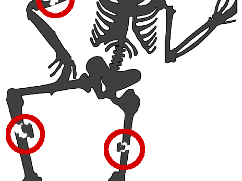
Osteosynthesis, the fixation of fractures by implants, is an important tool in orthopedics to facilitate the regeneration of bone and restore its functional behavior. The implant position and orientation after osteosynthesis is typically assessed visually based on several 2D X-ray images, showing the fixated bone from different angles. Due to the limited number of views and the perspective distortion in 2D X-rays, a visual assessment can, however, lead to erroneous conclusions about the actual success of the surgical outcome. In this project, we aim at utilizing computer-aided 3D reconstruction techniques to determine the 3D position of implants relative to the bone from post-operative 2D X-ray images. The purpose is not only to allow a quantitative assessment of the implant positioning for follow-up, but also to apply the reconstruction technique in large retrospective studies on osteosynthesis. Our goal is to gain insights on which position and orientation of the implant facilitates bone healing best.
3D-Reconstruction of femur and implant
Within the project, we are developing computer-assisted reconstruction methods to derive the 3D-positioning of fracture fixation implants w.r.t. the femur (thigh bone) based on plain 2D radiographs. The aim is to relate the shape of the bone and the implant in 3D space as depicted in post-operative X-rays, without a-priori knowledge about the patient-specific 3D shape of the femur. We, however, assume that the implant shape is given in terms of a computer-aided design (CAD) model, as for example provided by the implant manufacturer. In addition, the distance between the X-ray source and detector and the size of the detector used to generate the individual X-ray images need to be known. These X-ray parameters are usually standardized and stored digitally together with the 2D X-rays after screening.
The patient-specific shape and pose of the femur and the pose of the implant are estimated in three steps. First, the X-ray images are preprocessed in order to extract the outline of the implant/femur and to mask surrounding structures that are not reconstructed. In a second step, the known implant geometry is registered to each radiograph separately in 3D space. Based on the transformation of the implant in individual X-ray setups, a virtual setup is computed that relates the images spatially in a single coordinate system. The third step then consists of the 3D-reconstruction of the patient-specific femoral shape in the virtual X-ray setup. It is performed by means of an iterative process, in which a statistical shape and intensity model (SSIM) of the femur is fit to a pair of X-ray images until the model’s 2D projection onto the X-ray planes matches the anatomy depicted in the 2D references (see also 3D Reconstruction of Anatomical Structures from 2D X-ray Images).
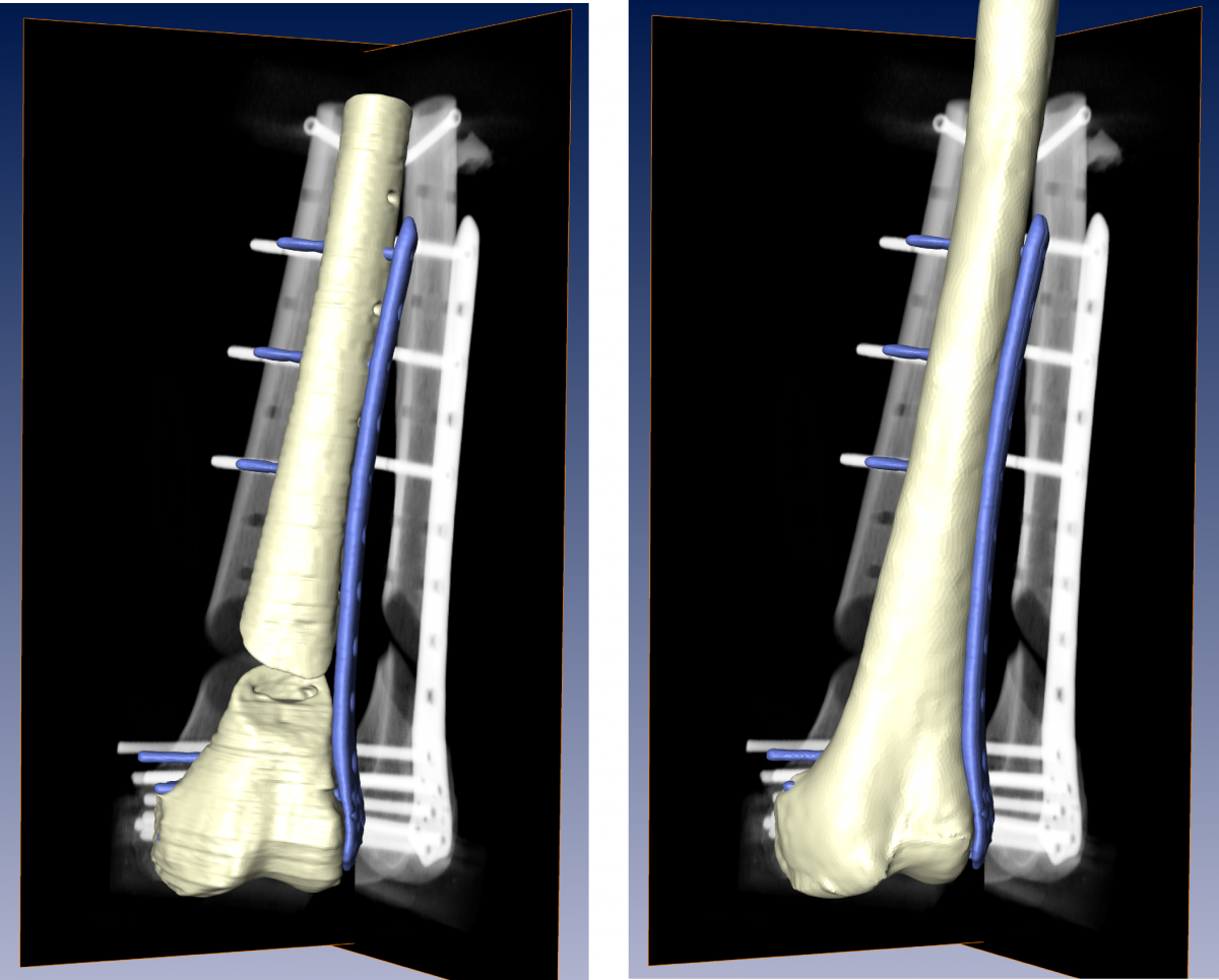
In a preliminary evaluation, we utilized the proposed method to derive the surface distance between implant plate and femur from 2D Digitally Reconstructed Radiographs (DRRs). A comparison to ground-truth from Computed Tomography shows that the the surface distance between plate and femur is reconstructed from 2D X-ray images with sub-millimeter accuracy on average.

Fig. 1. Ground-truth shape of the femur and implant from CT (left) and the 3D-reconstructed femoral shape and implant pose based on 2D X-ray images (right).
