This project aims at the development, analysis and implementation of algorithms for computer-assisted planning in hip surgery and hip joint replacement by fast virtual test. Fast forward simulations of patient-specific motion of hip joints and implants in 3D shall be enabled by exploiting suitable a priori information. To this end, we will derive, analyze, and implement reduced basis methods for heterogeneous joint models (reduced approximation).
Introduction
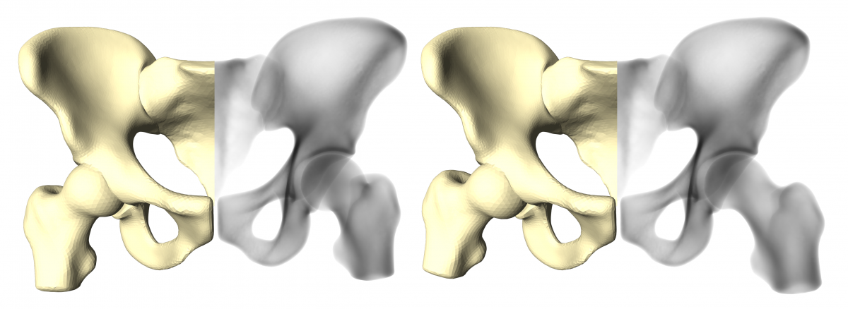
Statistical shape models (SSMs) prove to be an adequate prior in medical registration problems such as 3D segmentation of CT- and MRI data (Seim et al., 2008, see also Atlas-based 3D Image Segmentation). SSMs utilize the Principal Component Analysis (PCA) to express statistically significant anatomical variations within a training population. We enrich SSMs with volumetric bone-interior information learned from CT and utilize these Statistical Shape and Intensity Models (SSIMs) as a prior for the reconstruction process. A treatment plan in orthopaedics does often not only involve single bony anatomies, but rather whole joint structures (e.g. in total hip replacement). The individual components of the joint are oriented in various poses, depending on the patient's physical condition and pose in front of the X-ray scanner. To cope with different joint postures in the reference X-ray images, we further extend the SSIMs to articulated statistical shape and intensity models (ASSIMs). The ASSIM of the hip developed at ZIB represents the hip cup as a ball joint, in which each proximal femur (upper thigh bone) rotates with three degrees of freedom (see Fig. 1). The rotational center of the hip joint is part of the statistical analysis of the model and learned from the training population.

Fig. 1. Articulated Statistical Shape and Intensity Model (ASSIM) of the hip, expressing two different joint postures. Depicted here is the articulated surface of the model (beige) and the bone-interior density distribution (black and white).
Impingement Analysis
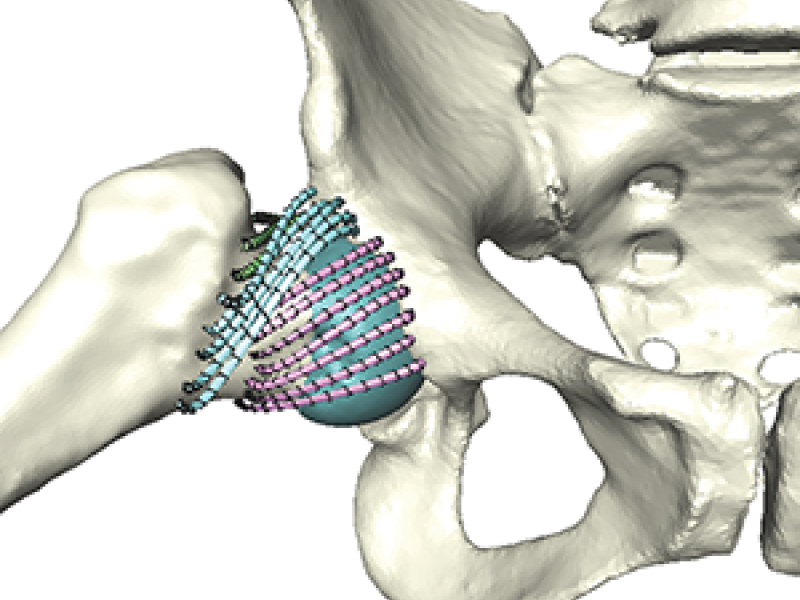
Upon moving the femur bones, possible collisions with other bone material, like the hip is detected and possible contact zones are displayed (see Fig. 2).

Fig. 2. Movement of femur bone of one hip instance. Once the transformation causes a collision, triangles on the surfaces are highlighted (center and right).
Collaboration
The project is done in collaboration with the "Numerics of Partial Differential Equations" group led by Dr. Ralf Kornhuber
