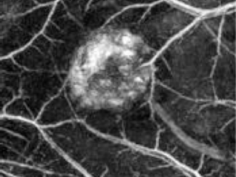
Photoacoustic (PA) imaging uses short optical pulses that are absorbed in tissue and cause ultrasound waves. The ultrasound waves are recorded, and reconstruction algorithms are then applied to compute a 3D image of the absorber distribution, such as hemoglobin. PA images that are acquired using visible or near-infrared light typically show the vasculature. PA tomography achieves a resolution of tens of μm at mm depths to hundreds of μm at cm depths. PA imaging provides strong contrast in vascularised soft tissues, where other modalities lack sensitivity. PA imaging has the potential for spatially resolved measurements of absolute chromophore concentrations and derived parameters, such as blood oxygen saturation. Deep tissue 3D quantitative photoacoustic tomography (qPAT), however, remains challenging due to the scale of the inverse problem and to date has not yet been demonstrated in vivo. The aim of this project is to deliver the first experimentally validated methodology for the non-invasive measurement of absolute blood oxygen saturation in deep tissue using photoacoustic tomography, which would be a major step in the translation of quantitative photoacoustic tomography towards a wide range of applications in the life sciences. The specific goals are to develop an efficient 3-D photoacoustic forward model; to develop computational and experimental methods for a tractable model-based inversion; and to validate practicable methods for the measurement of blood oxygen saturation in tissue phantoms and in vivo in a small animal model of tissue regeneration.
