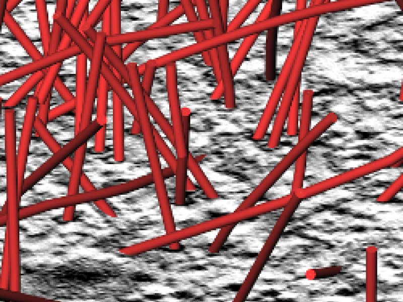
Microtubules are a part of the cytoskeleton of cells. They are tube-like polymeres that stabilize the cell but also play an important role in cell division. In particular the spindle - the microtubule assembly formed during cell division - is subject to intense research. One of the most important methods for looking at microtubule distribution in cells is using electron tomography. Here thick (300nm) sections of cells are prepared for electron microscopy and imaged on a CCD camera. Inverse radon transform algorithms are then used to reconstruct 3D information. One of the main bottle necks in the analysis of the microtubule network is then the construction of an exact model of the microtubules from the acquired data. This requires laborious manual modeling of microtubules. The aim of this project is to utilize image processing techniques to significantly accelerate the reconstruction of microtubules in the sections and thus reduce manual operator time and collect data in a way that is less biased by human operations.
Publications

2015
Reconstruction of Microtubule Centerlines from Electron Tomograms Doctoral thesis, Freie Universität Berlin, Knut Reinert (Advisor), 2015 Britta Weber BibTeX
Segmentation of Microtubules from Electron Tomograms
2014
Automated stitching of microtubule centerlines across serial electron tomograms PLoS ONE, p. e113222, 2014 (preprint available as ZIB-Report 14-41) Britta Weber, Erin M. Tranfield, Johanna L. Höög, Daniel Baum, Claude Antony, Tony Hyman, Jean-Marc Verbavatz, Steffen Prohaska PDF (ZIB-Report)
BibTeX
DOI
Segmentation of Microtubules from Electron Tomograms
The Segmentation of Microtubules in Electron Tomograms Using Amira Mitosis: Methods and Protocols, pp. 261-278, Springer, 2014 Stefanie Redemann, Britta Weber, Marit Möller, Jean-Marc Verbavatz, Anthony Hyman, Daniel Baum, Steffen Prohaska, Thomas Müller-Reichert BibTeX
DOI
Segmentation of Microtubules from Electron Tomograms
2012
Automated segmentation of electron tomograms for a quantitative description of actin filament networks Journal of Structural Biology, Vol.177, pp. 135-144, 2012 Alexander Rigort, David Günther, Reiner Hegerl, Daniel Baum, Britta Weber, Steffen Prohaska, Ohad Medalia, Wolfgang Baumeister, Hans-Christian Hege BibTeX
DOI
Segmentation of Microtubules from Electron Tomograms
Automated tracing of microtubules in electron tomograms of plastic embedded samples of Caenorhabditis elegans embryos Journal of Structural Biology, 178(2), pp. 129-138, 2012 Britta Weber, Garrett Greenan, Steffen Prohaska, Daniel Baum, Hans-Christian Hege, Thomas Müller-Reichert, Anthony Hyman, Jean-Marc Verbavatz BibTeX
DOI
Segmentation of Microtubules from Electron Tomograms
2011
Tuner: Principled Parameter Finding for Image Segmentation Algorithms Using Visual Response Surface Exploration IEEE Trans. Vis. Comput. Graph., 17(12), pp. 1892-1901, 2011 Thomas Torsney-Weir, Ahmed Saad, Torsten Möller, Hans-Christian Hege, Britta Weber, Jean-Marc Verbavatz BibTeX
Segmentation of Microtubules from Electron Tomograms
2009
Automatic alignment of stacks of filament data Proc. IEEE International Symposium on Biomedical Imaging, pp. 971-974, 2009 Vincent J. Dercksen, Britta Weber, David Günther, Marcel Oberlaender, Steffen Prohaska, Hans-Christian Hege PDF
BibTeX
Segmentation of Microtubules from Electron Tomograms