On the occasion of the international conference Visual Computing in Biology and Medicine (VCBM), which celebrated its 10th meeting this year, Hans-Christian Hege received a VCBM Award for his contribution to the development of the VCBM conference series and for his merits as a member of the steering committee.
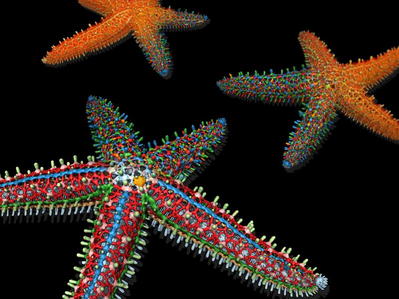
Additionally, the Best Image Award of the VCBM was received by a team consisting of members from ZIB, the Harvard University and the Max Planck Institute of Colloids and Interfaces (MPIKG). The team members — Daniel Baum and Hans-Christian Hege (ZIB), Lara Tomholt and James C. Weaver (Harvard University) and Mason N. Dean (MPIKG) — were honored for their image entitled “Dissecting a digital starfish".
Starfish and their relatives have an endoskeleton consisting of mineralized bones (ossicles). Among the species, a wide variety of osseous forms, connectivity and spatial organization of the ossicles occurs. The architecture of these skeletal elements plays a crucial role in species identification and the analysis of evolutionary relationships. The figure shows the three main steps in the reconstruction of the skeleton of a Pisaster giganteus from the temperate East Pacific. The starfish was imaged using micro computed tomography. Its endoskeletal elements (ossicles) were automatically segmented using a random-walk distance transformation and contour-tree segmentation. Finally the individual skeletal elements were classified and colored accordingly.