High-resolution synchrotron μCT allows one to study insects in a non-destructive way. This is particularly important for rare specimens. Despite the non-destructive nature of μCT, it still gives us information about the fine structure of the nervous system. However, the high resolution of the images as well as the complex anatomical structures of insects represent a challenge for segmenting and visualizing the specimens. In this project, we try to address these challenges, especially to support comparative visualization.
Approach
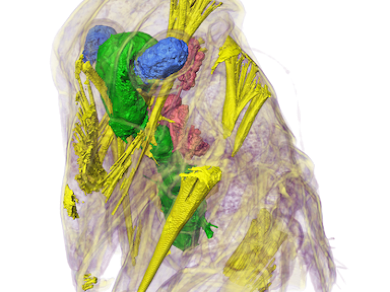
As a first approach we applied state-of-the-art segmentation tools like the seed-based watershed algorithm to segment detailed anatomical structures of the Hawaiian planthoppers Oliarus from high-resolution synchroton µCT images. A result can be seen in Fig. 1. The approach worked well but required a lot of user-interaction. A goal of this project will be to develop interactive segmentation tools that allows the biological expert to quickly segment anatomical structures. Furthermore, we aim at developing visualization tools that allow biologists to qualitatively as well as quantitatively compare anatomical structures of different species.

Fig 1.: Segmentation (from left to right): Manual seeds; result of seed-based watershed applied to the gradient-magnitude field of the image data; combination of surface rendering of segmented structures and volume rendering of µCT data set.
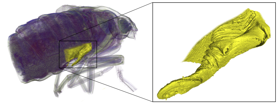
In a recent publication [Hoch, Wessel et al., 2014] we showed that high-resolution synchrotron µCT is well-suited to investigate even fine details of the lateral abdominal sensory and secretory organs (LASSO) of the taxon Bennini both in nymphs (Fig. 2) and adults.

Fig. 2: Segmentation of the LASSO of a Bennini nymph.
