One aim of this project is to analyze a large set of medical image data with respect to the anatomy of the knee joint. The aim is to determine the variation in shape of the knee and knee joint space, respectively bone and cartilage, between distal femur and proximal tibia. Clusters of similar shapes have to be determined in order to design a limited set of knee spacers that fit a wide range of the osteoarthritic population.

Data selection

In order to detect different clusters of similar shapes at least 500 MRI datasets need to be processed. The datasets are taken from The Osteoarthritis Initiative (OAI) database. The OAI database contains about 5000 patients. Therefore, a selection has been made based on the Kellgren-Lawrence OA-Score which is available for almost all patients. The Kellgren-Lawrence score differentiates between five grades. For each of the five grades 138 female and 145 male patients have been randomly selected resulting in a preselection of 1415 right knee MRI datasets. In a first step 500 of these datasets are processed and analysed. As MRI protocol SAG_3D_DESS_WE (sagittal 3D dual-echo steady state with selective water excitation) is used for bone and cartilage segmentation.

Data processing

Bone and cartilage of distal femur and proximal tibia are segmented automatically using linear Statistical Shape Models (SSMs) [1]. Errors in the automatic segmentation are corrected manually. Additionally, for each knee landmarks of the insertion sites of anterior cruciate ligament (ACL) and posterior cruciate ligament (PCL) are placed by hand. A schematic overview is given in figure 1.
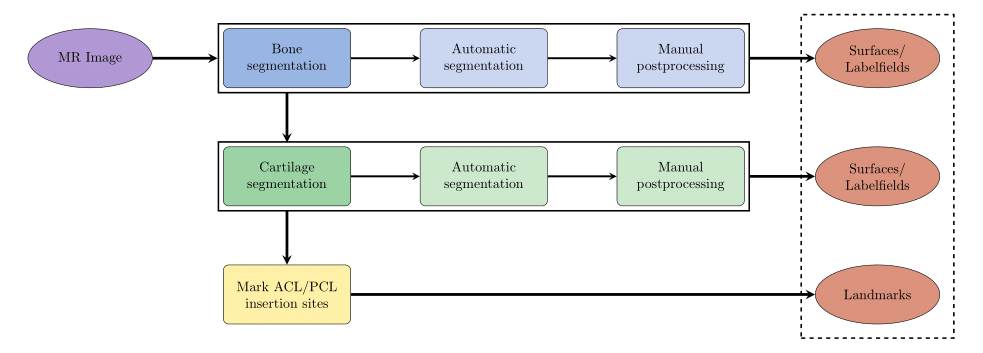
Fig. 1: Schematic overview of data processing

The following pictures illustrate the single steps of the pipeline.
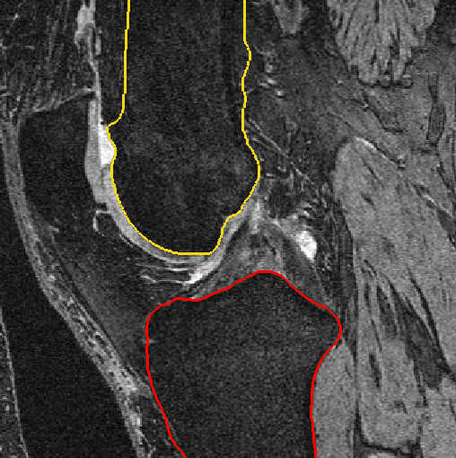
    

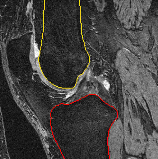
Fig. 2: Bone segmentation after automatic segmentation (left) and after manual postprocessing (right)
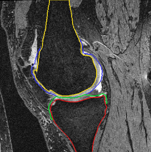
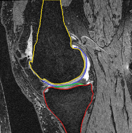
    

Fig. 3: Cartilage segmentation after automatic segmentation (left) and after manual postprocessing (right)
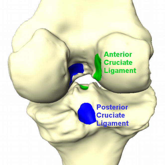
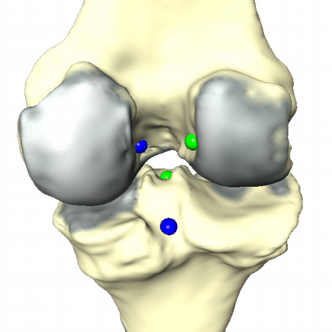
ACL_PCL_insertion_sites     Cartilage_interface_insertion_sites

Fig. 4: Insertion sites of ACL and PCL (left). Result of data processing (right). Cartilage thickness is displayed in red. ACL and PCL insertion sites are replaced by a representative point.


Analysis

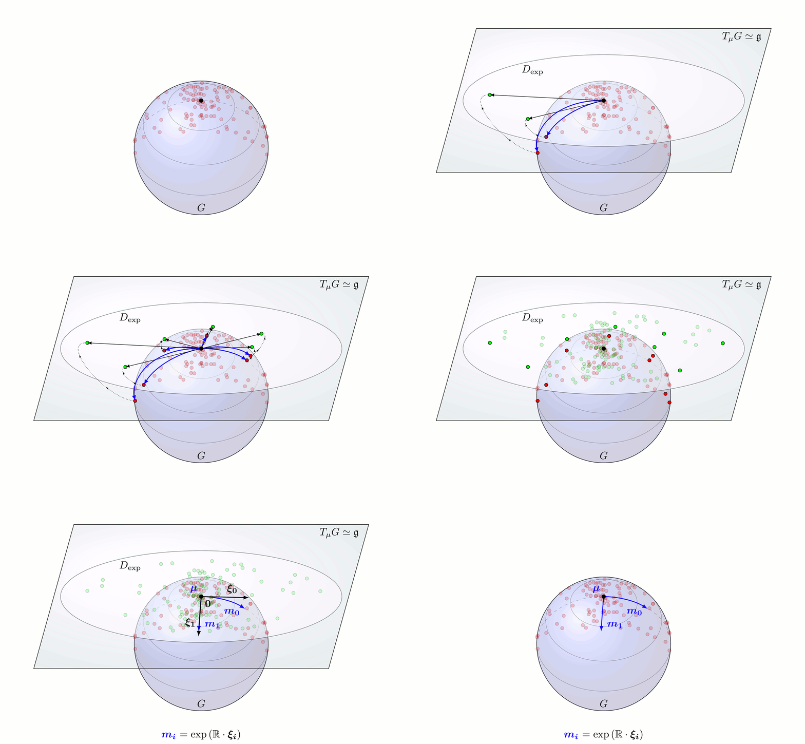
The aim is to find geometrical clusters in (very) high dimensional data. This fact complicates meaningful clustering. Therefore reduction of dimensionality/feature selection has to be done. Taking the geometries as triangular meshes there are different options how to proceed. One is the well known principal component analysis (PCA) on the vertex coordinates, that gives the SSMs, also known as Point Distribution Models (PDM). A more general approach is Principal Geodesic Analysis (PGA) [Ref Fletcher], which treats data as elements in a Riemannian manifold and carries out statistical operations therein or in the tangential space to a reference element respectively. This procedure has the advantage that it can be sensible to nonlinear shape changes depending on the chosen modeling space. It should be noted that PCA can be interpreted as special case of PGA, namely with the flat Euclidean space as underlaying manifold. We develop a method that employs the deformation gradient of a mesh modeled as element of a Lie group and thus delivering an even richer mathematical structure. It is called the Gradient Domain Model (GRM).
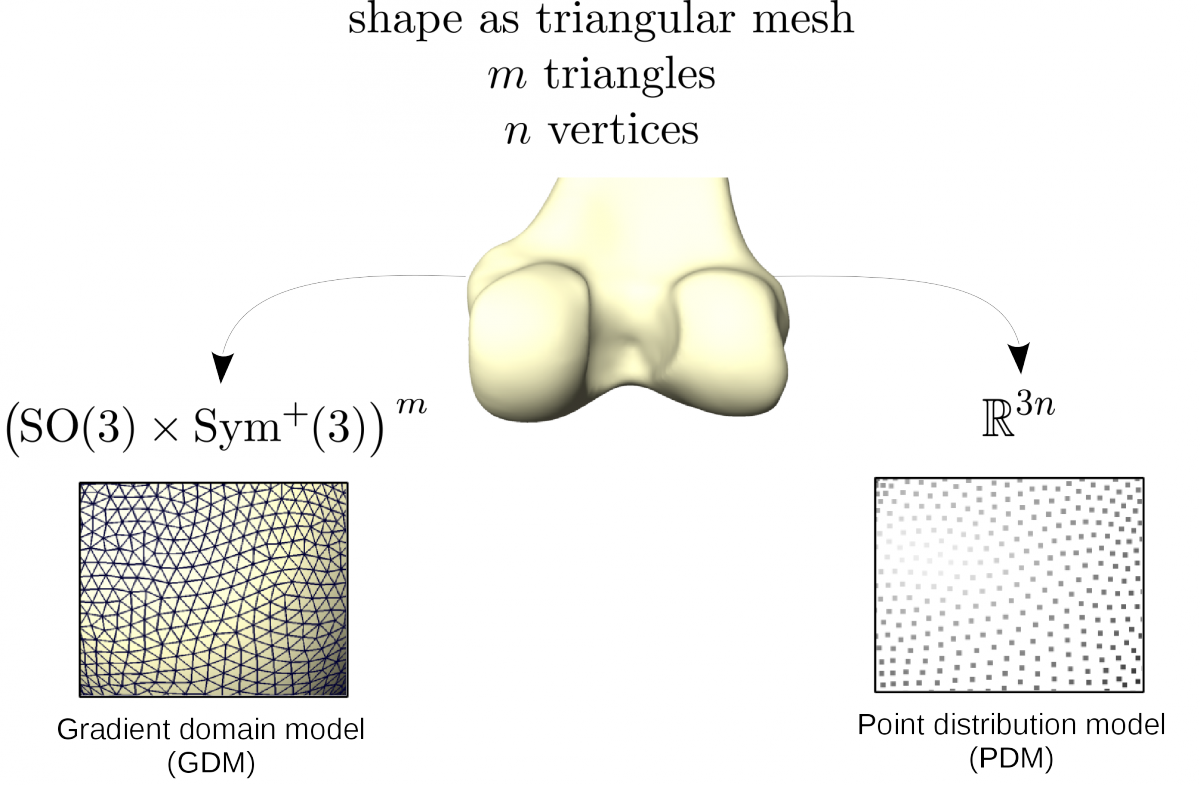
 

modelChoise

Fig. 5: Used properties of a mesh with respect to the chosen shape model.
 

pga_schematic 


Fig. 6: Principal Geodesic Analysis on a Lie group G schematically done on a sphere.


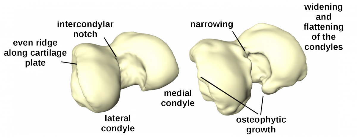
OA related changes of distal femur

The distal femur exibits clear pathological changes in case of a severe OA and is therefore an interesting structure to take into consideration for Clustering and Classification of OA.

KL0vsKL4  


Fig. 7: Healthy (left) and osteoarthritic (right) distal femur with delineated pathological changes in shape.


Classification and Clustering


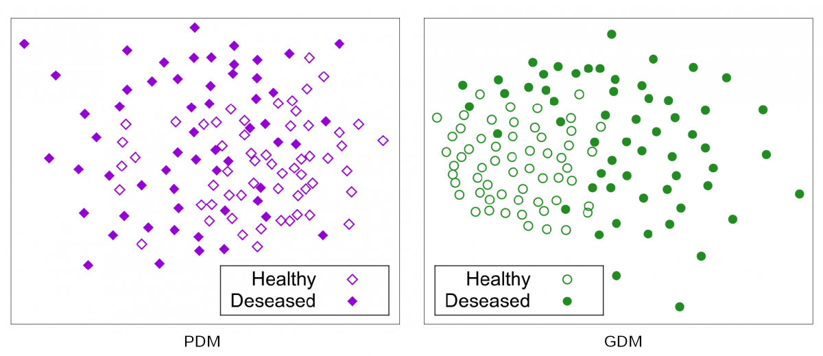
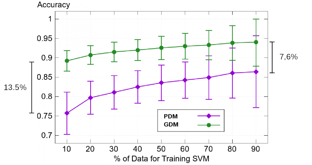
To find clusters we apply  our GDM on patients that are healthy (KL 0/1) and those that are severely deseased (KL 4). The resultant shape weights are used to train a Support Vector Machine in order to classify between this two groups.

class01


Fig. 8: Classification results for 58 severely deseased (KL4) and 58 (almost) healthy (KL0/1) femurs.
 

class02

Fig. 9: Sammon Projection for 58 severely deseased (KL4) and 58 (almost) healthy (KL0/1) femurs.


References

[1] Seim, H., Kainmueller, D., Lamecker, H., Bindernagel, M., Malinowski, J., & Zachow, S. (2010). Model-based auto-segmentation of knee bones and cartilage in MRI data. Proc. MICCAI Workshop Medical Image Analysis for the Clinic - A Grand Challenge, 215-223.


Publications

2022
Dynamic pressure analysis of novel interpositional knee spacer implants in 3D-printed human knee models Scientific Reports, Vol.12, 2022 Korbinian Glatzeder, Igor Komnik, Felix Ambellan, Stefan Zachow, Wolfgang Potthast PDF
BibTeX
DOI
Treating Osteoarthritis in Knee with Mimicked Interpositional Spacer
2021
Establishment of a rolling-sliding test bench to analyze abrasive wear propagation of different bearing materials for knee implants Applied Sciences, 11(4), 2021 Jessica Hembus, Felix Ambellan, Stefan Zachow, Rainer Bader BibTeX
DOI
Treating Osteoarthritis in Knee with Mimicked Interpositional Spacer
Towards novel osteoarthritis biomarkers: Multi-criteria evaluation of 46,996 segmented knee MRI data from the Osteoarthritis Initiative PLOS One, 16(10), 2021 Alexander Tack, Felix Ambellan, Stefan Zachow BibTeX
DOI
Treating Osteoarthritis in Knee with Mimicked Interpositional Spacer
2019
Automated Hip Knee Ankle Angle Determination using Convolutional Neural Networks Master's thesis, Otto-von-Guericke-Universität Magdeburg, Felix Ambellan, Alexander Tack, Stefan Zachow (Advisors), 2019 Henok Hagos Gidey PDF
BibTeX
URN
Treating Osteoarthritis in Knee with Mimicked Interpositional Spacer
Automated Segmentation of Knee Bone and Cartilage combining Statistical Shape Knowledge and Convolutional Neural Networks: Data from the Osteoarthritis Initiative Medical Image Analysis, 52(2), pp. 109-118, 2019 (preprint available as ZIB-Report 19-06) Felix Ambellan, Alexander Tack, Moritz Ehlke, Stefan Zachow PDF (ZIB-Report)
BibTeX
DOI
Treating Osteoarthritis in Knee with Mimicked Interpositional Spacer
Automated Segmentation of Knee Bone and Cartilage combining Statistical Shape Knowledge and Convolutional Neural Networks: Data from the Osteoarthritis Initiative (Supplementary Material) Medical Image Analysis, 52(2), pp. 109-118, 2019 (OAI-ZIB dataset) Felix Ambellan, Alexander Tack, Moritz Ehlke, Stefan Zachow BibTeX
DOI
Treating Osteoarthritis in Knee with Mimicked Interpositional Spacer
Comparison of 2D and 3D CNNs for Classification of Knee MRI Bachelor's thesis, Freie Universität Berlin, Alexander Tack, Felix Ambellan (Advisors), 2019 Mona Prendke PDF
BibTeX
URN
Treating Osteoarthritis in Knee with Mimicked Interpositional Spacer
Statistical Shape Models - Understanding and Mastering Variation in Anatomy Biomedical Visualisation, Paul M. Rea (Ed.), Springer Nature Switzerland AG, 1, pp. 67-84, 2019, ISBN: 978-3-030-19384-3, ISBN: 978-3-030-19385-0 (preprint available as ZIB-Report 19-13) Felix Ambellan, Hans Lamecker, Christoph von Tycowicz, Stefan Zachow PDF (ZIB-Report)
BibTeX
DOI
Treating Osteoarthritis in Knee with Mimicked Interpositional Spacer
2018
An Efficient Riemannian Statistical Shape Model using Differential Coordinates Medical Image Analysis, 43(1), pp. 1-9, 2018 (preprint available as ZIB-Report 16-69) Christoph von Tycowicz, Felix Ambellan, Anirban Mukhopadhyay, Stefan Zachow PDF (ZIB-Report)
BibTeX
DOI
Treating Osteoarthritis in Knee with Mimicked Interpositional Spacer
Automated Segmentation of Knee Bone and Cartilage combining Statistical Shape Knowledge and Convolutional Neural Networks: Data from the Osteoarthritis Initiative Medical Imaging with Deep Learning, 2018 Felix Ambellan, Alexander Tack, Moritz Ehlke, Stefan Zachow PDF
BibTeX
Treating Osteoarthritis in Knee with Mimicked Interpositional Spacer
Changes in Knee Shape and Geometry Resulting from Total Knee Arthroplasty Journal of Engineering in Medicine, 232(1), pp. 67-79, 2018 Mohsen Akbari Shandiz, Paul Boulos, Stefan Sævarsson, Heiko Ramm, Chun Kit Fu, Stephen Miller, Stefan Zachow, Carolyn Anglin BibTeX
DOI
Treating Osteoarthritis in Knee with Mimicked Interpositional Spacer
2017
Computerassistierte Auswahl und Platzierung von interpositionalen Spacern zur Behandlung früher Gonarthrose Proceedings of the Jahrestagung der Deutschen Gesellschaft für Computer- und Roboterassistierte Chirurgie (CURAC), pp. 106-111, Vol.16, 2017 (preprint available as ZIB-Report 17-72) Robert Joachimsky, Felix Ambellan, Stefan Zachow PDF (ZIB-Report)
BibTeX
URN
Treating Osteoarthritis in Knee with Mimicked Interpositional Spacer
Evaluating two methods for Geometry Reconstruction from Sparse Surgical Navigation Data Proceedings of the Jahrestagung der Deutschen Gesellschaft für Computer- und Roboterassistierte Chirurgie (CURAC), pp. 24-30, Vol.16, 2017 (preprint available as ZIB-Report 17-71) Felix Ambellan, Alexander Tack, Dave Wilson, Carolyn Anglin, Hans Lamecker, Stefan Zachow PDF (ZIB-Report)
BibTeX
URN
Treating Osteoarthritis in Knee with Mimicked Interpositional Spacer
Validation of three-dimensional models of the distal femur created from surgical navigation point cloud data for intraoperative and postoperative analysis of total knee arthroplasty International Journal of Computer Assisted Radiology and Surgery, 12(12), pp. 2097-2105, 2017 David Wilson, Carolyn Anglin, Felix Ambellan, Carl Martin Grewe, Alexander Tack, Hans Lamecker, Michael Dunbar, Stefan Zachow PDF
BibTeX
DOI
Treating Osteoarthritis in Knee with Mimicked Interpositional Spacer
2016
Statistical Shape Modeling of Musculoskeletal Structures and Its Applications Computational Radiology for Orthopaedic Interventions, Springer, pp. 1-23, 2016, ISBN: 978-3-319-23481-6 Hans Lamecker, Stefan Zachow BibTeX
DOI
Treating Osteoarthritis in Knee with Mimicked Interpositional Spacer