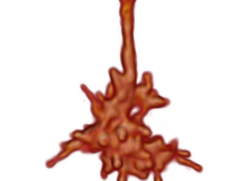
2-Photon microscopy has opened a rapidly expanding field of imaging studies in intact tissues and living animals. Diverse lymphatic organs, kidney, heart, skin, and brain can be examined. 2-Photon microscopy enables studies about intact nervous systems and investigation of growth cone dynamics.
Tracking and quantification of neuronal filopodia dynamics in 3D 2-photon microscopy
Filopodia dynamics are thought to control growth cone guidance during brain wiring, but the types and roles of growth cone dynamics underlying neuronal circuit assembly in a living brain are largely unknown. To address this issue, a brain culture live-image system that is applicable for all developmental stages of Drosophila brain was developed. The used 2-photon microscope allows generating 3D time series of R7 growth cones of different age, genetic manipulation, or chemical treatment. To analyze and compare the filopodia dynamics in the acquired datasets of different types, an efficient method for information extraction and quantification is required. We developed a semi-automatic pipeline in the 3D Software Amira that allows 3D reconstruction and tracking of filopodia represented as skeleton graphs. Those graphs simplify the extraction of statistical information like filopodial length, number, orientation, retraction, and extensions.
Analysis of Vesicle Trafficking in Axons
The dynamics of transport vesicles during neural development are still unknown. Vesicles can transport proteins and metabolic substances from the soma to the axon terminal and back. We track and analyze vesicles in time-lapse 2-photon microscopy by building Markov sate models based on trajectories. The stationary distribution and committor probabilities along the axon clearly indicate retrogade transport.
