This research project investigates the improvement of root canal treatment (RCT) by analyzing the relationship between root canal morphologies and treatment failures. This pursuit of enhancing the longevity and success of root canal treatments is the central motivation driving our project.
Project goals
There is a clear clinical need to improve the outcomes of root canal treatments, yet current 2D clinical data like x-rays are often insufficient to thoroughly analyze potential causes of treatment failure. To address this gap, we acquire high-resolution 3D micro-CT scans, which allow us to investigate the complex relationships between root canal morphology, procedural or material-related defects, and the behavior of different dental materials.These detailed 3D datasets enable us to formulate hypotheses about how procedural, anatomical and material-related factors may influence treatment success. Our long-term goal is to translate this knowledge into a clinically applicable decision support system based on conventional dental X-rays. Ultimately, our project aims to contribute to improved treatment techniques and protocols, better-suited instrumentation, and standardized methods for evaluating new filling and sealing materials.
Latest Development
We have developed two complementary tools designed for the efficient analysis of root canal treatment (RCT) defects within high-resolution micro-CT data. The first is an automated segmentation pipeline that handles complex, high-throughput data processing to isolate key tooth structures. The second is Segplorer, an interactive 3D visualization and analysis toolkit that allows researchers to precisely detect, interact with, and quantitatively characterize structural defects such as pores and delaminations. Together, these tools bridge the gap between massive raw imaging datasets and actionable, quantitative research insights.
(1) A high-throughput pipeline for segmenting high-resolution 3D (HR3D) micro-CT datasets.
(2) Segplorer: A toolkit for Interactive 3-D visualization and quantitative characterization of pores and delaminations.
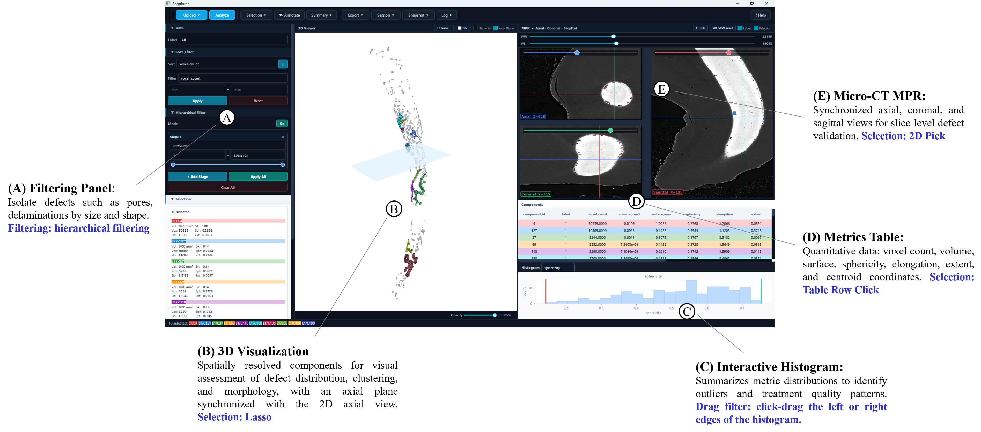
Segplorer treats RCT defects as 3D components to quantify their size, shape, and distribution within the sealer interzone. By synchronizing 3D rendering, multiplanar 2D views, and interactive filtering/histograms, it replaces tedious manual workflows with an efficient, reproducible framework for exploring complex defect patterns.
Project Overview
DFG-Forschergruppe 2804 "InterDent"
The DFG-funded research group "InterDent" aims to enhance collaboration among materials, computer, and dental sciences. Our goal is to reduce uncertainty about what defines a 'durable dental interzone' in RCT by obtaining high-resolution, standardized data and integrating insights from advanced computational methods. The following animation presents a demo created with our Amira software, showcasing a micro-CT scan along with deep learning-based segmentation of various tooth structures.

Project description
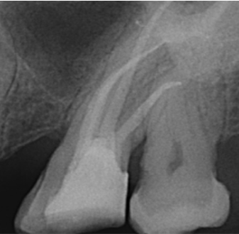
(1) Dental x-ray of a treated tooth:
Dental x-rays are commonly used in RCT for pre-operative planning and post-operative assessment, but their limited resolution hinders the detection and analysis of treatment defects, such as gaps or voids in the sealer interzone, and they do not provide a 3D view of the root canal system.
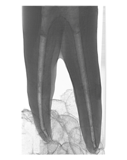
(2) Micro-CT of a treated tooth:
Micro-CT, conducted in a laboratory setting using extracted tooth samples, offers ultra-high-resolution imaging of the root canal system, enabling precise differentiation of dental materials. Micro-CT is therefore exceptionally well-suited for 3D quantitative analysis of root canal morphology. However, this high resolution comes at a cost: each individual scan can generate 50–75 GB of data. The resulting data volume imposes significant challenges for algorithm development, particularly for tasks like automatic segmentation.
Managing, processing, and analyzing such large datasets requires substantial computational power, memory, and storage infrastructure, making the development and deployment of efficient computational methods particularly demanding. In the meantime, we are also developing a standardized platform for collecting and sharing large micro-CT datasets. This platform will integrate our tools for automatic segmentation and failure analysis, providing valuable support to both dental practitioners and researchers.
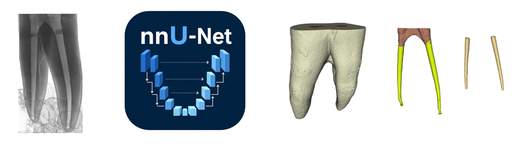
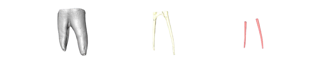
(3) Root canal segmentation from Micro-CT:
Reliable morphological analysis of the root canal system depends on accurate segmentation of high-resolution dental imaging (e.g., micro-CT), where treatment defects are clearly visible. We manually annotated selected slices of a micro-CT scan, and trained a deep learning based segmentation model. Then, we applied the segmentation model to the rest of the slices to automatically segment these tooth structures.
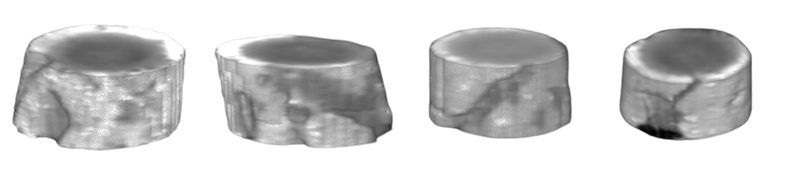
(4) Defect analysis:
Once we have an accurate segmentation of the tooth structures, especially the sealer inter-zone, we can detect the defects within the region of interest. From there, we can perform subsequent analysis, e.g., classifying them into different types of defects, like pore, delamination and etc, as well as analyzing the correlation between their occurrence and the morphology of the sealer inter-zone.
(5) Translation of knowledge from high-resolution micro-CT to x-rays in clinical routine:
Even though ideal for morphological analysis, micro-CT is not suitable for use in clinical routine. Transferring the knowledge of defect occurrence gained from 3D morphological analysis of the root canal system enabled by micro-CT to routine dental imaging, such as x-rays, is highly desirable.
Our partners