In the course of the 350 million years (Lower Devonian-Upper Cretaceous) of ammonoid evolution, the iterative trend of development starting with simple to complexly, folded septa of the phragmocone is well documented. Changes in the ratio of chamber volume against the inner chamber surface allow statements about different lifestyles in different ontogenetic stages and comparisons between different taxa.
Recent enhancements in tomographic imaging techniques enable a non-invasive view inside fossil structures with astonishing spatial resolution. However, a manual segmentation to reconstruct the structure of fossil ammonites from tomographic data is very labor intensive and time consuming, requiring several weeks for one ammonoid specimen. In case the signal to noise ratio (SNR) is low or imaging artifacts occur, an automatic reconstruction process turns out to be too difficult to derive useful information from the tomography data using current techniques.
Our first aim is therefore to develop specialized algorithms for image denoising. We are implementing a multi-level approach which runs on highly parallelized hardware such as graphic boards (GPU). Further, a concept will be developed which allows fast, automated segmentation of CT-data and a subsequent morphological analysis – not only in cross-section but also showing three-dimensional developmental patterns during ontogeny.
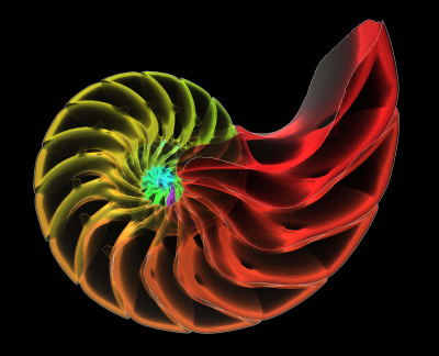
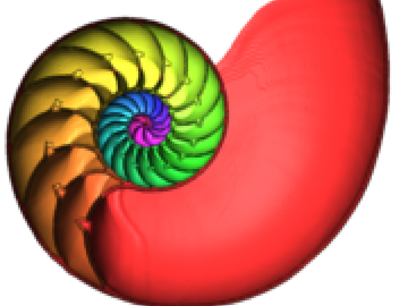
The shell of the recent Nautilus pompilius, which already inspired D´Arcy Thompson for his book "On growth and form" (1917).
Introduction
The analysis of the shapes of fossils, either fully or partially preserved within the surrounding rocks, requires imaging methods that are primarily used in non-destructive testing. Current state-of-art enhanced tomographic imaging techniques enable a view inside of solid objects with astonishing spatial resolution. This processing of tomographic image data is commonly used in medicine, where typical tasks are image processing (filtering, enhancement), segmentation (classification of structures), and geometric reconstruction of segmented structures.
The aim is to develop specialized algorithms for image denoising taking advantage of self-similarity within the image data to being able to reconstruct small structures from noisy image data. Therefore, variants of non-local-means (NLM) filtering techniques are to be investigated with respect to their suitability for our kind of data. In addition we are planning to investigate whether NLM algorithms (for denoising) can be combined with nonlinear anisotropic diffusion (NAD) algorithms for feature enhancement and extraction as it has been introduced for noise reduction in electron tomography images.
Being able to geometrically reconstruct septal surfaces with sufficient accuracy, the visualization and the quantitative analysis of such structures will be another goal. Tools are needed to reduce the amount of data and to measure volumes, areas, or other measures of interest such as ratios between surface areas and volumes.
Scope
The syn-apomorphic development of a hydrostatic apparatus with a chambered shell and water mass regulation by a siphuncle system is an essential basic pattern characteristic of cephalopods. The design of septa and their specific attachment on the inside whorl (i.e., the suture line) is therefore of essential functional and phylogenetic significance for ammonoids.
The geometric reconstruction of anatomical structures from tomographic image data is well established in medical diagnosis and therapy planning (Zachow et al., 2007). However, while in medical imaging the image resolution is between 0.25 and 1mm, which in most cases is sufficient for diagnostic purposes, this resolution is by far not sufficient for the reconstruction or quantitative assessment of the subjects of study (ammonite septa and juvenile ammonite shell), whose finest structures are between 5 and 20 micro meters. Image processing in medicine is not particularly focused on denoising of the image data but rather on automation of the segmentation process in order to avoid error prone, labor intensive manual delineation of structures within the image data.
Ammonite volumetric datasets obtained from micro-CT images show a low signal-to-noise ratio (SNR) and multiple imaging artifacts. This makes the reconstruction process too difficult to derive useful information from the image data using available methods in medical imaging. Due to the very thin septa of the juvenile stage of ammonoids, common methods of image filtering for noise reduction are insufficient for our purpose. Another handicap is the size of the data sets. Tomographic imaging with high resolution often leads to data sets of several tenths of GB that usually cannot processed with standard computer hardware.
A manual segmentation process is very labor intensive and time consuming, requiring several weeks for one ammonoid specimen. Thus, enhanced image processing algorithms are needed to filter image data in such a way that SNR is improved while inherent structures are preserved. With the development of new filter algorithms, as well as fully automated or at least semi-automatic segmentation methods, the classification of the structures of interests (ammonoid septa, siphuncle) and the work flow will be significantly improved.
Material and data acquisition
Smaller quantities of suitable material from north-eastern Germany (Aptian / Albian local glacial erratic boulders) were acquired (Fig. 1). Other localities with adequate preservation were found in Kamchatka (Russia), Hokkaido (Japan) and north of Moscow (Kostroma/Unzha area). From the Callovian (middle Jurassic) of the Kostroma area a small amount of suitable material (mainly kosmoceratids, cadoceratids and perisphinctids) is at hand and available for our project.
A quantifiable geometrical and functional-analysis has failed so far due to the low scanning resolution of 50 µm in commercially available CT-scanners. Due to the well known sampling theorem the spatial resolution of the scanning system needs to be at least twice as high as the smallest structure that needs to be reconstructed from the image data. Therefore special scanning devices are required which are operated in maximum resolution mode. The Steinmann Institute (Rheinische Friedrich Wilhelms Universität Bonn, working group of Prof. T. Martin) has already given the permission to use the micro-CT device v|tome|x s (GE phoenix|x-ray) for a fee. With a first test a resolution of about 10 µm was achieved with this device.
However, the maximum resolution mode itself already produces a very low signal-to-noise ratio (SNR) due to high sensitivity and sensor artifacts. In result, so called noise is degrading the image quality, thus hindering automatic segmentation algorithms to distinguish between relevant structures of interest and noise.
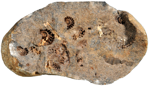
Fig. 1) Hollow ammonites Leymeriella in a Gault concretion from north-eastern Germany (coll. J. Kalbe).
Previous work
At Zuse Institute Berlin (ZIB) a system is being developed that provides algorithms to reconstruct geometry from 3D image data. This software, which is also commercially available, is constantly improved with respect to research problems.
Our first tests have been performed on different data sets to assess the requirements for imaging and 3D reconstruction (Fig. 2). It became obvious that new algorithms for specific image processing tasks have to be developed that allow for a faithful 3D reconstruction of thin structures such as septal surfaces and suture lines in ammonoids.
Further tests have proven that only the very rare preservation of ammonites in hollow condition is suitable for the creation of digital 3D-computer models using Amira. Ammonites with the chambers filled with sediment, secondary calcite or pyrite are unsuitable for such studies due to complete x-ray absorption with shadowing artifact (Fig. 3).
Using manual and semi-automatic tools we were able to segment the complete shell of an ammonite including septa and juvenile shell parts in a very time-consuming process. Our aim is to develop specialized algorithms for image denoising taking advantage of self-similarity within the image data to being able to reconstruct small or thin structures from noisy image data.
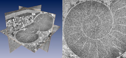
Fig. 2) Multiplanar reconstruction (MPR) of a tomographic dataset of 9 GB, acquired with a v|tome|x s (180kV tube current). MPR and visualisation with ZIBAmira on a Quad Core PC with 24 GB RAM, running Windows 7 Ultimate. Left) Complete data set loaded into memory. Right) detail that clearly depicts image noise due to bad signal to noise ratio.
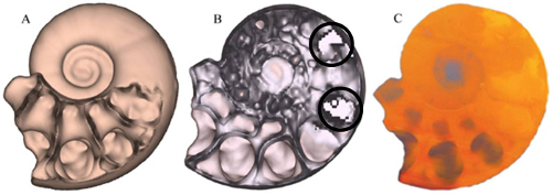
Fig. 3) CT-scan of Argonauticeras besairei. A) first chambers filled with spherulitic calcite covering the walls which caused a distinct kind of smoothening-effect, B) the earlier chambers showing pyrite which absorbs most of the x-rays and inhibits the visualisation process (white pixelated areas), C) in the volume rendering scalar values of the 3D image are integrated in the direction of the projection producing a transparent image, that shows the sedimentary infill of nearly all of the earlier chambers. Due to similar density the sediment has a similar colour code compared to the aragonitic shell, that makes both undistinguishable for reconstruction (coll. H. Keupp).
Fast non-local-means denoising
Preliminary results
The non-local-means (NLM) algorithm tries to take advantage of the high degree of redundancy of any natural image, where every small patch has many similar patches in the same image. The NLM algorithm computes the value of every single pixel as a weighted sum of all pixels in the image, where pixels whose surrounding neighborhoods are similar to the current pixel in the original image are assigned higher weights.
Naïve implementations of this method are too slow, and in our case, with high-resolution datasets containings thousands of millions of volumetric data elements, are not feasible. Approaches to the problem of high-dimensional filtering can be found that share two common features:
- Use of principal component analysis (PCA) to reduce the dimensionality of the input data (in our case, the space of all possible fixed-size patches that can be generated from the volumetric data).
- Use of special data structures to reduce the number of samples and approximate the required computations.
We selected the NLM approach based on adaptive manifolds. As first step we evaluated the quality of the results that could be obtained from the Ammonite dataset. We adapted the current implementation to support volumetric data, and proceeded to generate different results exploring the parameter space. To speedup this search, we used different small subvolumes extracted from the original dataset (Fig. 4). We used a single-threaded implementation in CPU, and each test required between 5 and 15 minutes of computation.
After fine-tuning the parameters, we tried an automatic segmentation test by applying a sobel filter on the volume, and then a simple thresholding (Fig. 5). Even for this simple segmentation, important portions of the septa and the shell can be retrieved.
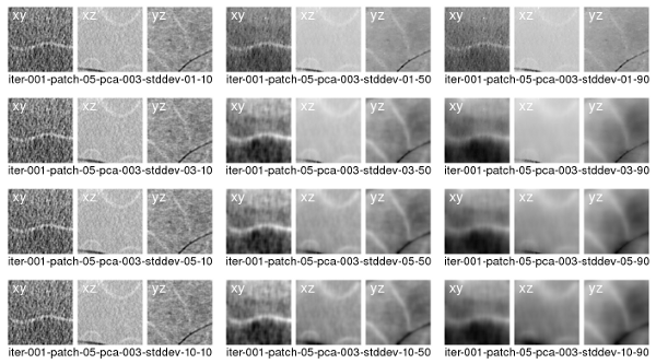
Fig. 4) Results from denosing a small subvolume from the Ammonite dataset using NLM. For each result, different values of spatial and range standard deviation were chosen. For each result, three slices oriented in different directions are shown.
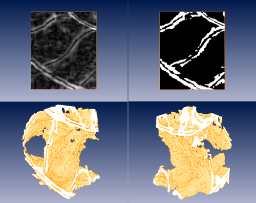
Fig. 5) Top, from left to right: result of a sobel edge detector on the denoised dataset; segmentation of septa and shell material by thresholding on the result of the edge detector. Bottom: two views of volume rendering of the previous segmentation, where portions of the septa and the siphuncle can be clearly identified.
